

| Journal of Neurology Research, ISSN 1923-2845 print, 1923-2853 online, Open Access |
| Article copyright, the authors; Journal compilation copyright, J Neurol Res and Elmer Press Inc |
| Journal website http://www.neurores.org |
Original Article
Volume 9, Number 3, June 2019, pages 28-34
Repetitive Transcranial Magnetic Stimulation in Treatment of Levodopa-Induced Dyskinesia in Parkinson’s Disease
Aktham Ismail Alemama, c, Mohamed Abdelhalim Eltantawib
aDepartment of Neuropsychiatry, Faculty of Medicine, Menoufia University, Menoufia, Egypt
bDepartment of Neurology, Mansoura New General Hospital, Dakahlia, Egypt
cCorresponding Author: Aktham Ismail Alemam, Department of Neuropsychiatry, Faculty of Medicine, Menoufia University, Menoufia, Egypt
Manuscript submitted November 14, 2018, accepted January 3, 2019
Short title: rTMS for LID in PD Patients
doi: https://doi.org/10.14740/jnr512
| Abstract | ▴Top |
Background: Dyskinesia is one of the major complications of long-term dopaminergic treatment of Parkinson’s disease (PD), and deep brain stimulation may be the only satisfactory treatment for it. This study aims to search if there is any therapeutic effect of repetitive transcranial magnetic stimulation (rTMS) for levodopa-induced dyskinesia (LID) in PD patients.
Methods: This study was conducted in Mansoura International Specialized Hospital in 43 complicated idiopathic PD patients. The patients with LID were divided into two groups matched in age, sex, duration and stage of the disease. Dyskinesia was detected by the Unified PD Rating Scale section IV. The patients of the study group (20 patients) received active rTMS, 5 Hz was applied bilaterally over the motor hand and leg areas of the cortex, in 20 trains, and each train is formed of 100 pulses, with 20-s inter-train interval. Ten sessions were administered once per day for 10 successive days for each patient. The patients of the control group (20 patients) received sham rTMS.
Results: After rTMS, there was significant improvement in LID in the study group (P < 0.001), while there was no improvement in the control one (P = 0.585). As regards to the LID clinical presentations, there was no significant difference between the two groups according to dyskinesia duration (P = 0.246), disability (P = 0.425) and early morning dystonia (P = 0.059), while there was significant improvement of painful dyskinesia in the study group (P = 0.046).
Conclusions: The rTMS may have an additional therapeutic benefit for LID. Further studies may put the best rTMS parameters to establish more prominent and longer-lasting clinical effects.
Keywords: Parkinson’s disease; rTMS; Dyskinesia
| Introduction | ▴Top |
The incidence of Parkinson’s disease (PD) has been estimated to be 4.5 - 21 cases per 100,000 population per year, and most studies yield a prevalence of approximately 120 cases per 100,000 population [1]. In Egypt the crude prevalence rate of PD was 557/100,000 [2]. Dyskinesia is one of the real complications of long haul dopaminergic treatment of PD. The pathophysiology and the clinical risk factors causing dyskinesia in PD are not completely comprehended, and their medical treatment is still inadequate [3, 4]. The revealed incidence rates of levodopa-induced dyskinesia (LID) demonstrate a wide range, from 9-80% [5]. Deep brain stimulation surgery, which is affirmed by Food and Drug Administration for the intractable complications emerging from long-standing dopaminergic treatment [6], does not improve them in numerous patients with PD [7]. Patients with LID have three main clinical presentations: peak dose dyskinesia, wearing-off or off-period dystonia, and diphasic dyskinesia. The peak dose dyskinesia is the most widely recognized and diphasic dyskinesia is the least common [8]. The response to therapy in PD is affected by the age of onset and severity of PD, dose and duration of levodopa (L-dopa) treatment, low body weight, L-dopa/carbidopa/entacapone treatment, female gender, higher Unified PD Rating Scale (UPDRS) score, and some genetic polymorphisms [9]. Some factors that affect both pre- and post-synaptic nigrostriatal dopamine transmission may prompt these motor complications [10].
Transcranial magnetic stimulation (TMS) was first introduced by Barker et al, who scientifically proved the influence of magnetic stimulation on motor cortex of the human brain in 1985 [11].
Repetitive TMS (rTMS) is an easy and noninvasive procedure of brain stimulation in view of the hypothesis of electromagnetic induction. A review of the literature, including 2,228 patients, revealed that rTMS does not carry significant risk of adverse events in the PD population [12]. It is the repetitious application of TMS pulses over a predefined target with ability to alter excitability at the site of stimulation of the brain [13], and it also affects brain areas anatomically connected to the stimulation site [14]. The rTMS at frequencies of 5 Hz and higher noteworthily increases motor cortex excitability, while the rTMS at frequencies of 1 Hz and lower depresses it [15]. It was found that in normal people, use of rTMS over the primary motor area (M1) [16] or the dorsolateral prefrontal cortex (DLPFC) [17] provoked ipsilateral dopamine discharge from the putamen and caudate respectively. During the last two decades, rTMS has been inspected as a possible treatment for PD [18]. Also, rTMS may help in improving some complications of PD as it has a positive effect in on-freezers with advanced PD with subsequent decrease of number of falls [19].
The outcomes are clashing, and no reasonable treatment protocol has yet been characterized. Although some of the researches that used high-frequency studies in the range of 25 Hz demonstrated positive improvements [20], the study that used theta burst stimulation (50 Hz) failed to exhibit this effect [21].
The aim of our study was to search if there is any helpful therapeutic impact of rTMS on LID in PD patients.
| Patients and Methods | ▴Top |
This study was conducted from May 2016 to May 2017, in Mansoura International Specialized Hospital in 43 patients with idiopathic PD complicated by dyskinesia. The study protocol has been approved by the institute’s committee on human research. All patients fulfilled the UK PD brain bank criteria. All patients subjected to the study were right handed and above 50 years old with the presence of at least two of the three cardinal signs (tremors, bradykinesia and rigidity) [22]. They showed strong dramatic response to L-dopa initially, and then the response started to wane with appearance of motor complications. All patients were complaining of LID. It included chorea, choreoathetosis, or dystonia [23].
The exclusion criteria included history of seizures, previous cerebral strokes, severe head trauma, anti-psychotic drugs intake, substance or alcohol abuse, signs of serious cognitive impairment (Mini-Mental State Examination score < 24), substance or alcohol abuse, deep brain stimulation, direct current stimulation or electro-convulsive therapy. The patients with secondary parkinsonism and parkinsonian plus syndromes were excluded.
Proper history taking of general and neurological complaints and full neurological examination were done. The UPDRS is used to follow the longitudinal course of PD. It is made up of six sections: 1) Part I: evaluation of mentation, behavior, and mood; 2) Part II: self-evaluation of the activities of daily life (ADLs) including speech, swallowing, handwriting, dressing, hygiene, falling, salivating, turning in bed, walking, and cutting food; 3) Part III: clinician-scored monitored motor evaluation; 4) Part IV: complications of therapy; 5) Part V: Hoehn and Yahr staging of severity of PD; and 6) Part VI: Schwab and England ADL scale [22]. We used UPDRS section IV to assess the duration of dyskinesia, and associated disability and pain.
The duration of dyskinesia was calculated as: what proportion of the waking day was dyskinesia present (historical information)? The grades were: 0 = none, 1 = 1-25% of day, 2 = 26-50% of day, 3 = 51-75% of day and 4 = 76-100% of day.
The disability was evaluated as: how disabling was the dyskinesia (historical information)? The grades were: 0 = not disabling, 1 = mildly disabling, 2 = moderately disabling, 3 = severely disabling, and 4 = completely disabled.
Painful dyskinesia was estimated as: how painful was the dyskinesia? The grades were: 0 = no painful dyskinesias, 1 = slight, 2 = moderate, 3 = severe and 4 = marked.
The presence of early morning dystonia was taken from the historical information and reported as: 0 = no and 1 = yes.
In this randomized, sham-controlled study, the 43 patients were divided into a study group and a control one. The study group included initially 23 patients. The patients received active rTMS, 5 Hz bilaterally over the motor hand and leg areas of the cortex. All patients were on both L-dopa and anticholinergic drugs for more than 5 years. All of them were on a fixed dose of their anti-parkinsonian medication for at least 1 month before starting the study until the end of the study. The doses varied between three and four tablets daily of L-dopa/carbidopa 250/25 mg in each tablet. No anti-dyskinetic medications were given before, during, or after TMS treatment. Three patients could not tolerate the pain under the coil and so they withdrew from the study. The control group comprises 20 complicated PD patients who received sham rTMS. It is matched with the study group as regards to the age, sex, duration, and staging of the disease. We chose sham stimulation for the control arm to offset the potential placebo effect of the rTMS intervention. The procedure was explained to both groups and written informed consent was taken after approval by the local ethical committee.
During the session, the patients were seated comfortably in a reclining chair. The biphasic rTMS pulses were delivered through circular coil (outer diameter = 14 cm; maximum field strength = 1.9 Tesla) attached to MagPro R30 stimulator (MagVenture, Farum, Denmark) (Fig. 1). TMS pulses were delivered through the coil which is positioned perpendicular to the central sulcus line. The study group received 5 Hz applied in 20 trains, and each train is formed of 100 pulses, with 20-s inter-train interval. Ten sessions were administered once per day for 10 successive days for each patient. The control group could not tell which type of stimulation was active or sham. The position of the sham coil was identical to that of the active coil. The look and feel of the sham stimulation were very similar but not identical to active stimulation. They incorrectly picked sham stimulation as active.
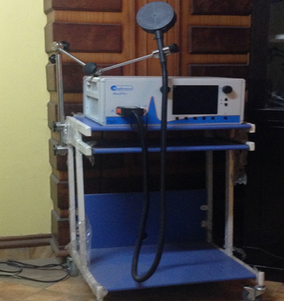 Click for large image | Figure 1. MagPro R30 magnetic stimulator (MagVenture, Farum, Denmark, 2012). |
Statistical analysis
Data was analyzed using Statistical Package for Social Sciences (SPSS) version 15 (SPSS Inc., Chicago, IL, USA). Qualitative data was presented as number and percent. Comparison between groups was done by Chi-square test. Quantitative data was presented as mean ± SD. Paired t-test was used for comparison within groups. The P < 0.05 was considered statistically significant. Student t-test was used to compare between two groups.
| Results | ▴Top |
Forty PD patients who complicated with LID have completed the study, and they were divided into two groups: study group with 20 patients who were subjected to active rTMS, and the control group with 20 patients who were subjected to sham rTMS.
In the study group, nine patients were men and 11 patients were women. Their age ranged between 60 and 75 years (66.65 ± 4.58). Their illness duration ranged between 62 - 80 (68.10 ± 5.35) months. In the control group, 12 patients were men and eight patients were women. Their age ranged between 58 and 78 years (66.60 ± 5.97). Their illness duration ranged between 63 - 80 (68.75 ± 5.30) months (Table 1).
 Click to view | Table 1. Comparison Between Demographic Data of the Study and Control Group Patients Before rTMS |
Before stimulation, the study group was matched in every aspect to the control one. There was no significant difference in age, sex or duration of the disease. As regards to the clinical staging of the disease before rTMS, there was no statistical difference between both groups (Table 2).
Click to view | Table 2. Comparison Between Both Groups as Regards to the Disease Staging Before rTMS |
According to the individual clinical presentations of patients with LID, active stimulation did not lead to much improvement regarding dyskinesia duration (P = 0.246), disability (P = 0.45) or early morning dystonia (P = 0.059) at the end of the study (Table 3), while it showed significant improvement regarding painful dyskinesia (P = 0.046) (Table 4).
Click to view | Table 3. The Comparison Between Clinical Presentations of LID in Study and Control Groups Before rTMS |
Click to view | Table 4. Comparison Between Individual Clinical Presentations of LID in Study and Control Groups After rTMS |
The results showed significant improvement in LID (as a whole involving the total score of the four clinical parameters) in the study group at the end of active rTMS, but no improvement in the control group after sham stimulation (Table 5).
Click to view | Table 5. Comparison Between Dyskinesia Before and After rTMS in Each Group |
| Discussion | ▴Top |
Some controlled and uncontrolled researches have investigated the therapeutic use of rTMS in PD and have discovered advantageous impacts [24]. The results of our study about the effect of rTMS on LID in patients with PD showed significant improvement (P < 0.001). It did not show much improvement regarding dyskinesia duration, disability or early morning dystonia, while it showed significant improvement regarding painful dyskinesia (P = 0.046).
Our outcomes were in concurrence with Filipovic et al [25], who led a randomized controlled examination on 10 patients with severe LID using real and sham rTMS (1,800 pulses; 1-Hz rate more than 4 days). Despite the fact that the study and control groups reacted in same extent, just the study group showed significant changes toward the end of treatment. The stimulation protocol and the overall dose used in the study may be too low to give significant differences between the study and control groups. Also, Brusa et al demonstrated that a low-recurrence (rTMS) to the supplementary motor area (SMA) diminished LID yet just for up to 30 min. In their consequent study, they conveyed stimulation for 5 continuous days (every day sessions for 15 min), but there was no cumulative improvement developed toward the end of treatment [26].
Similarly, our results were in the same direction of Elahi et al [18], who found improvement of dyskinesia that went on for around 30 min when they conveyed a low-frequency rTMS (1-Hz frequency, 900 stimuli over 15 min) over the SMA.
Additionally, Wagle Shukla et al [27] used the parameters of 900 stimuli at 1 Hz more than 15 min for 2 weeks, and focused on the primary motor cortex rather (M1) than the SMA. They discovered significant improvement at 1 day and 2 weeks evaluation in the dyskinesia rating scale and in the scores based off a diary maintained by patients.
Additionally, Rascol et al, who performed an investigation of 10 day by day sessions of low frequency rTMS over motor cortex, indicated reduction of peak dose dyskinesia, which was quantifiable and huge a day after the last session [28]. As dyskinesia in PD by most accounts may be due to over-activity in M1, this element of low frequency rTMS may well be behind the watched clinical benefit in this study.
On the other hand, Wagle Shukla et al [27] found that sham rTMS induced significant placebo effect on LID in some patients, and even in few cases, the sham rTMS effect was greater than the effect of active rTMS. These results raise the possibility that the reduction in dyskinesia scores observed following the active rTMS may be due to placebo effect rather than a genuine rTMS effect.
Also, a meta-analysis of pooled results from 10 controlled trials found that there was a therapeutic effect for the use of high-frequency rTMS, while there was no significant effect for low-frequency rTMS studies [29].
The differences in the outcomes of different studies might be because of the varieties in sample sizes, and rTMS dosing regimens, result measures, inclusion and exclusion criteria, use of sham TMS, brain side and sites of stimulation, medication use, disease stage, age, sex and side of onset of PD.
Compared with other rTMS protocols used for treatment of patients with PD, we used circular coil for stimulation. This produced diffuse effect in the nearby cortical areas. We stimulated the site of M1 bilaterally similar to Khedr et al [30]. Regarding number of sessions we applied 10 sessions, while others as Arias et al [29] and Benninger et al [31] applied seven sessions, Bornke et al [32] and Koch et al [33] applied single sessions. As regards frequency we used 5 Hz, while others used frequency of 10 Hz or higher [34].
Ying-hui et al found that there were no significant differences in effect size between rTMS sites (M1 versus other frontal regions) and between high-frequency and low-frequency rTMS. However, there was a significant difference in effect size among different combinations of rTMS frequency and rTMS site. The effect sizes estimated from high-frequency rTMS targeting M1 and from low-frequency rTMS applied over other frontal regions were significant. The effect sizes obtained from the other two combinations of rTMS frequency and rTMS site were not significant [35].
The pathophysiology of LID may be related to the striatal degeneration that depends on the dose and duration of L-dopa treatment [36, 37]. Presynaptic disturbances generate large dopamine oscillation in the brain and postsynaptic changes leading to abnormal responses in dopaminergic neurons. Progressive degeneration of presynaptic nigral neurons results in loss of dopamine storage capacity causing sudden rise in dopamine level leading to peak-dose dyskinesia and sudden decline in dopamine level causing wearing-off dyskinesia [38]. A critical number of patients do not create dyskinesia in spite of prolonged L-dopa treatment. Hereditary variables could assume a part in deciding the event of peak-dose dyskinesia. Certain kinds of hereditary polymorphism of the dopamine receptor D2 quality have been related with a lessened danger of creating peak-dose dyskinesia [39].
Intermittent intake of L-dopa results in downstream changes in proteins and genes, causing the changes in the striatal output that induce the dyskinesia [40]. Disinhibition of the primary and associated motor cortex secondary to increased outflow (pallidothalamocortical motor pathway) may be responsible for LID [26].
A few other nondopaminergic frameworks including glutamatergic, gamma-aminobutyric, serotonergic, histaminergic, adenosine, and cannabinoid receptors have been hypothesized to assume a vital part in the advancement of LID [41].
The literature on the mechanism of action of rTMS for treatment of LID is scanty [42, 43]. Some studies demonstrated a long-lasting beneficial effect of low-frequency rTMS targeted at the SMA. The increased levels of activity in these frontal regions may result in the inhibition of action via a hyperdirect pathway that connects several frontal regions (including the SMA, DLPFC, and inferior frontal gyrus) and the subthalamic nucleus [44]. A specific impairment in plasticity of the cortical inhibitory mechanisms was demonstrated in PD patients with dyskinesia [45]. The benefits of rTMS in treatment of LID are sustained at a follow-up period of about 6 weeks [20].
Our study found that rTMS resulted in a statistically significant improvement in painful dyskinesia but not in the other three measures. TMS acts on pain-modulating systems in the diencephalon and descending pain pathways from the brainstem to the spinal cord [46]. Several studies proposed hypotheses for the mechanism of chronic pain management with TMS. GABAergic inhibitory neurotransmission may play a role in pain modulation with TMS [47]. Endogenous opioid systems may also be affected by TMS-induced analgesia depending on the stimulation site [48]. One of the suggested mechanisms is the efficacy of rTMS in treating depression which is a common co-morbidity in chronic pain [49].
One of the limitations of this study is the small number of cases. Also, although the UPDRS is helpful in assessment of different aspects of dyskinesia, it does not include the anatomical distribution of dyskinesia in different body parts [50]. Additionally, the rTMS in this study was delivered with patients on medication. It is yet unknown whether the effects may be different and even stronger if rTMS is delivered off medication. But when therapeutic effect of rTMS was assessed in many trials [31] during the off state and in others [30] during the on state, the estimated effect sizes were not significantly different between the off-state and on-state evaluations.
The encouraging results of this study, together with the cumulative results from other researches, suggest a need for further studies that would systematically evaluate relevant methodological features (e.g. more days of rTMS, stronger TMS pulses, bilateral stimulation, rTMS in on versus off medication) to get more accurate protocols that may lead to more prominent and longer-lasting clinical improvements in LID. Other studies may evaluate the effect of rTMS on the non-motor manifestations of PD, for example, the cognitive functions which are affected early in the disease [51], or fatigue and sleep disturbances [52], and may evaluate the effect of rTMS on the non-dopaminergic extra-striatal sites that may be involved in PD as the centromedian-parafasicular complex of the thalamus [53].
Conclusions
The rTMS showed therapeutic effect on LID in PD. Although the improvement was not significant regarding dyskinesia duration, disability or early morning dystonia, it was significant regarding painful dyskinesia.
Informed Consent
Subjects have given their informed consent.
| References | ▴Top |
- Muangpaisan W, Mathews A, Hori H, Seidel D. A systematic review of the worldwide prevalence and incidence of Parkinson's disease. J Med Assoc Thai. 2011;94(6):749-755.
- Khedr EM, Al Attar GS, Kandil MR, Kamel NF, Abo Elfetoh N, Ahmed MA. Epidemiological study and clinical profile of Parkinson's disease in the Assiut Governorate, Egypt: a community-based study. Neuroepidemiology. 2012;38(3):154-163.
doi pubmed - Baas H. Dyskinesia in Parkinson's disease. Pathophysiology and clinical risk factors. J Neurol. 2000;247(Suppl 4):IV/12-16.
doi - Fabbrini G, Brotchie JM, Grandas F, Nomoto M, Goetz CG. Levodopa-induced dyskinesia. Mov Disord. 2007;22:1379-1389.
doi pubmed - Impact of deprenyl and tocopherol treatment on Parkinson's disease in DATATOP patients requiring levodopa. Parkinson Study Group. Ann Neurol. 1996;39(1):37-45.
doi pubmed - Okun MS, Gallo BV, Mandybur G, Jagid J, Foote KD, Revilla FJ, Alterman R, et al. Subthalamic deep brain stimulation with a constant-current device in Parkinson's disease: an open-label randomised controlled trial. Lancet Neurol. 2012;11(2):140-149.
doi - Wagle Shukla A, Okun MS. Surgical treatment of Parkinson's disease: patients, targets, devices, and approaches. Neurotherapeutics. 2014;11(1):47-59.
doi pubmed - Vijayakumar D, Jankovic J. Drug-Induced Dyskinesia, Part 1: Treatment of Levodopa-Induced Dyskinesia. Drugs. 2016;76(7):759-777.
doi pubmed - Warren Olanow C, Kieburtz K, Rascol O, Poewe W, Schapira AH, Emre M, Nissinen H, et al. Factors predictive of the development of Levodopa-induced dyskinesia and wearing-off in Parkinson's disease. Mov Disord. 2013;28(8):1064-1071.
doi pubmed - Cenci MA. Presynaptic Mechanisms of l-DOPA-Induced Dyskinesia: The Findings, the Debate, and the Therapeutic Implications. Front Neurol. 2014;5:242.
doi pubmed - Barker AT, Jalinous R, Freeston IL. Non-invasive magnetic stimulation of human motor cortex. Lancet. 1985;1(8437):1106-1107.
doi - Vonloh M, Chen R, Kluger B. Safety of transcranial magnetic stimulation in Parkinson's disease: a review of the literature. Parkinsonism Relat Disord. 2013;19(6):573-585.
doi pubmed - Wagle Shukla A, Vaillancourt DE. Treatment and physiology in Parkinson's disease and dystonia: using transcranial magnetic stimulation to uncover the mechanisms of action. Curr Neurol Neurosci Rep. 2014;14(6):449.
doi pubmed - Reithler J, Peters JC, Sack AT. Multimodal transcranial magnetic stimulation: using concurrent neuroimaging to reveal the neural network dynamics of noninvasive brain stimulation. Prog Neurobiol. 2011;94(2):149-165.
doi pubmed - Pascual-Leone A, Valls-Sole J, Wassermann EM, Hallett M. Responses to rapid-rate transcranial magnetic stimulation of the human motor cortex. Brain. 1994;117( Pt 4):847-858.
doi pubmed - Fukuda M, Edwards C, Eidelberg D. Functional brain networks in Parkinson's disease. Parkinsonism Relat Disord. 2001;8(2):91-94.
doi - Strafella AP, Paus T, Fraraccio M, Dagher A. Striatal dopamine release induced by repetitive transcranial magnetic stimulation of the human motor cortex. Brain. 2003;126(Pt 12):2609-2615.
doi pubmed - Elahi B, Elahi B, Chen R. Effect of transcranial magnetic stimulation on Parkinson motor function - systematic review of controlled clinical trials. Mov Disord. 2009;24(3):357-363.
doi pubmed - Hatem Shehata, Nevin Shalaby, Mohamed El-Tamawy, Shaimaa Jaafary. rTMS for On-Freezers with Advanced PD. A long term follow up study. Egyptian Journal of Neurology, Psychiatry & Neurosurgery. 2013;50(4):355-360.
- Lomarev MP, Kanchana S, Bara-Jimenez W, Iyer M, Wassermann EM, Hallett M. Placebo-controlled study of rTMS for the treatment of Parkinson's disease. Mov Disord. 2006;21(3):325-331.
doi pubmed - Benninger DH, Berman BD, Houdayer E, Pal N, Luckenbaugh DA, Schneider L, Miranda S, et al. Intermittent theta-burst transcranial magnetic stimulation for treatment of Parkinson disease. Neurology. 2011;76(7):601-609.
doi pubmed - Fahn S, Elton RL. UPDRS Development Committee. Unified Parkinson's disease Rating Scale. In: Fahn Marsden CD, Clane DB, Goldstein M, eds, Recent developments in Parkinson's disease. Florham Park, New Jersey: Macmillan. 1987; p. 153-163.
- Fahn S. The spectrum of levodopa-induced dyskinesias. Ann Neurol. 2000;47(4 Suppl 1):S2-9; discussion S9-11.
- Fregni F, Simon DK, Wu A, Pascual-Leone A. Non-invasive brain stimulation for Parkinson's disease: a systematic review and meta-analysis of the literature. J Neurol Neurosurg Psychiatry. 2005;76(12):1614-1623.
doi pubmed - Filipovic SR, Rothwell JC, van de Warrenburg BP, Bhatia K. Repetitive transcranial magnetic stimulation for levodopa-induced dyskinesias in Parkinson's disease. Mov Disord. 2009;24(2):246-253.
doi pubmed - Brusa L, Versace V, Koch G, Iani C, Stanzione P, Bernardi G, Centonze D. Low frequency rTMS of the SMA transiently ameliorates peak-dose LID in Parkinson's disease. Clin Neurophysiol. 2006;117(9):1917-1921.
doi pubmed - Wagle-Shukla A, Angel MJ, Zadikoff C, Enjati M, Gunraj C, Lang AE, Chen R. Low-frequency repetitive transcranial magnetic stimulation for treatment of levodopa-induced dyskinesias. Neurology. 2007;68(9):704-705.
doi pubmed - Rascol O, Sabatini U, Brefel C, Fabre N, Rai S, Senard JM, Celsis P, et al. Cortical motor overactivation in parkinsonian patients with L-dopa-induced peak-dose dyskinesia. Brain. 1998;121( Pt 3):527-533.
doi pubmed - Arias P, Vivas J, Grieve KL, Cudeiro J. Controlled trial on the effect of 10 days low-frequency repetitive transcranial magnetic stimulation (rTMS) on motor signs in Parkinson's disease. Mov Disord. 2010;25(12):1830-1838.
doi pubmed - Khedr EM, Rothwell JC, Shawky OA, Ahmed MA, Hamdy A. Effect of daily repetitive transcranial magnetic stimulation on motor performance in Parkinson's disease. Mov Disord. 2006;21(12):2201-2205.
doi pubmed - Benninger DH, Iseki K, Kranick S, Luckenbaugh DA, Houdayer E, Hallett M. Controlled study of 50-Hz repetitive transcranial magnetic stimulation for the treatment of Parkinson disease. Neurorehabil Neural Repair. 2012;26(9):1096-1105.
doi pubmed - Bornke C, Schulte T, Przuntek H, Muller T. Clinical effects of repetitive transcranial magnetic stimulation versus acute levodopa challenge in Parkinson's disease. J Neural Transm Suppl. 2004;68:61-67.
doi - Koch G, Brusa L, Caltagirone C, Peppe A, Oliveri M, Stanzione P, Centonze D. rTMS of supplementary motor area modulates therapy-induced dyskinesias in Parkinson disease. Neurology. 2005;65(4):623-625.
doi pubmed - Shirota Y, Ohtsu H, Hamada M, Enomoto H, Ugawa Y, Research Committee on r TMSToPsD. Supplementary motor area stimulation for Parkinson disease: a randomized controlled study. Neurology. 2013;80(15):1400-1405.
doi pubmed - Chou YH, Hickey PT, Sundman M, Song AW, Chen NK. Effects of repetitive transcranial magnetic stimulation on motor symptoms in Parkinson disease: a systematic review and meta-analysis. JAMA Neurol. 2015;72(4):432-440.
doi pubmed - Jenner P. Molecular mechanisms of L-DOPA-induced dyskinesia. Nat Rev Neurosci. 2008;9(9):665-677.
doi pubmed - Iravani MM, McCreary AC, Jenner P. Striatal plasticity in Parkinson's disease and L-dopa induced dyskinesia. Parkinsonism Relat Disord. 2012;18(Suppl 1):S123-125.
doi - Metman LV, Konitsiotis S, Chase TN. Pathophysiology of motor response complications in Parkinson's disease: hypotheses on the why, where, and what. Mov Disord. 2000;15(1):3-8.
doi - Oliveri RL, Annesi G, Zappia M, Civitelli D, Montesanti R, Branca D, Nicoletti G, et al. Dopamine D2 receptor gene polymorphism and the risk of levodopa-induced dyskinesias in PD. Neurology. 1999;53(7):1425-1430.
doi pubmed - Bibbiani F, Costantini LC, Patel R, Chase TN. Continuous dopaminergic stimulation reduces risk of motor complications in parkinsonian primates. Exp Neurol. 2005;192(1):73-78.
doi pubmed - Chase TN, Bibbiani F, Oh JD. Striatal glutamatergic mechanisms and extrapyramidal movement disorders. Neurotox Res. 2003;5(1-2):139-146.
doi pubmed - Lefaucheur JP, Drouot X, Von Raison F, Menard-Lefaucheur I, Cesaro P, Nguyen JP. Improvement of motor performance and modulation of cortical excitability by repetitive transcranial magnetic stimulation of the motor cortex in Parkinson's disease. Clin Neurophysiol. 2004;115(11):2530-2541.
doi pubmed - Dragasevic N, Potrebic A, Damjanovic A, Stefanova E, Kostic VS. Therapeutic efficacy of bilateral prefrontal slow repetitive transcranial magnetic stimulation in depressed patients with Parkinson's disease: an open study. Mov Disord. 2002;17(3):528-532.
doi pubmed - Nambu A, Tokuno H, Inase M, Takada M. Corticosubthalamic input zones from forelimb representations of the dorsal and ventral divisions of the premotor cortex in the macaque monkey: comparison with the input zones from the primary motor cortex and the supplementary motor area. Neurosci Lett. 1997;239(1):13-16.
doi - Morgante F, Espay AJ, Gunraj C, Lang AE, Chen R. Motor cortex plasticity in Parkinson's disease and levodopa-induced dyskinesias. Brain. 2006;129(Pt 4):1059-1069.
doi pubmed - Lefaucheur JP. Use of repetitive transcranial magnetic stimulation in pain relief. Expert Rev Neurother. 2008;8(5):799-808.
doi pubmed - Barr MS, Farzan F, Davis KD, Fitzgerald PB, Daskalakis ZJ. Measuring GABAergic inhibitory activity with TMS-EEG and its potential clinical application for chronic pain. J Neuroimmune Pharmacol. 2013;8(3):535-546.
doi pubmed - de Andrade DC, Mhalla A, Adam F, Texeira MJ, Bouhassira D. Neuropharmacological basis of rTMS-induced analgesia: the role of endogenous opioids. Pain. 2011;152(2):320-326.
doi pubmed - Loo CK, Mitchell PB. A review of the efficacy of transcranial magnetic stimulation (TMS) treatment for depression, and current and future strategies to optimize efficacy. J Affect Disord. 2005;88(3):255-267.
doi pubmed - Goetz CG, Tilley BC, Shaftman SR, Stebbins GT, Fahn S, Martinez-Martin P, Poewe W, et al. Movement Disorder Society-sponsored revision of the Unified Parkinson's Disease Rating Scale (MDS-UPDRS): scale presentation and clinimetric testing results. Mov Disord. 2008;23(15):2129-2170.
doi pubmed - Al Serafy OA, Soliman RH, Asaad RE, Tawfik MH. Cognitive functions in patients with Parkinson's disease: II. Impact of genetic factors. Egyptian Journal of Neurology, Psychiatry and Neurosurgery. 2015;52(1):29-36.
- Shalash AS, Hamid E, Elrassas HH, Bedair AS, Abushouk AI, Khamis M, Hashim M, et al. Non-motor symptoms as predictors of quality of life in egyptian patients with Parkinson's disease: a cross-sectional study using a culturally adapted 39-item Parkinson's disease questionnaire. Front Neurol. 2018;9:357.
doi pubmed - Alemam AI, Obeid OF. Additional Extra-striatal Site of Neurodegeneration in Parkinson's Disease. Egyptian Journal of Neurology, Psychiatry & Neurosurgery. 2013;50(3):221-225.
This article is distributed under the terms of the Creative Commons Attribution Non-Commercial 4.0 International License, which permits unrestricted non-commercial use, distribution, and reproduction in any medium, provided the original work is properly cited.
Journal of Neurology Research is published by Elmer Press Inc.