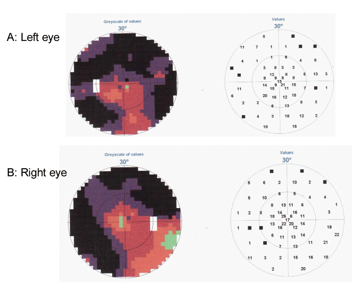
Figure 1. Perimetry test demonstrating loss of peripheral vision on Left eye (A) and Right eye (B).
| Journal of Neurology Research, ISSN 1923-2845 print, 1923-2853 online, Open Access |
| Article copyright, the authors; Journal compilation copyright, J Neurol Res and Elmer Press Inc |
| Journal website http://www.neurores.org |
Case Report
Volume 2, Number 2, April 2012, pages 54-58
Blindness or Agnosia: Review of Posterior Cortical Atrophy and a Difficult Case
Figures
Table
| 2009 | 2010 | |
|---|---|---|
| WAIS: Wechsler Adult Intelligence Scale; WMS: Wechsler Memory Scale; *Not examined. | ||
| WAIS-III Vocabulary | 20/66 point | * |
| Digit span - forward | 5 | 4 |
| Digit span - backward | 0 | 0 |
| Sentence repetition | 13/22 | 10/22 |
| Selective Reminding Test (Buschke) Immediate recall | 58/100 correct | 67/100 correct |
| Selective Reminding Test - Delayed recall | 5/10 correct | 7/10 correct |
| Selective Reminding Test - Recognition | 8/10 correct | 8/10 correct |
| WMS-III Logical Memory I | 7/75 | * |
| WMS-III Logical Memory II | 4/50 | * |