Journal of Neurology Research, ISSN 1923-2845 print, 1923-2853 online, Open Access
Article copyright, the authors; Journal compilation copyright, J Neurol Res and Elmer Press Inc
Journal website http://www.neurores.org

Case Report

Volume 2, Number 1, February 2012, pages 25-29


Botulinum Toxin Injection Into the Cricopharyngeal Muscle for Syringobulbia-induced Dysphagia: A Case Report

Figures

Figure 1.
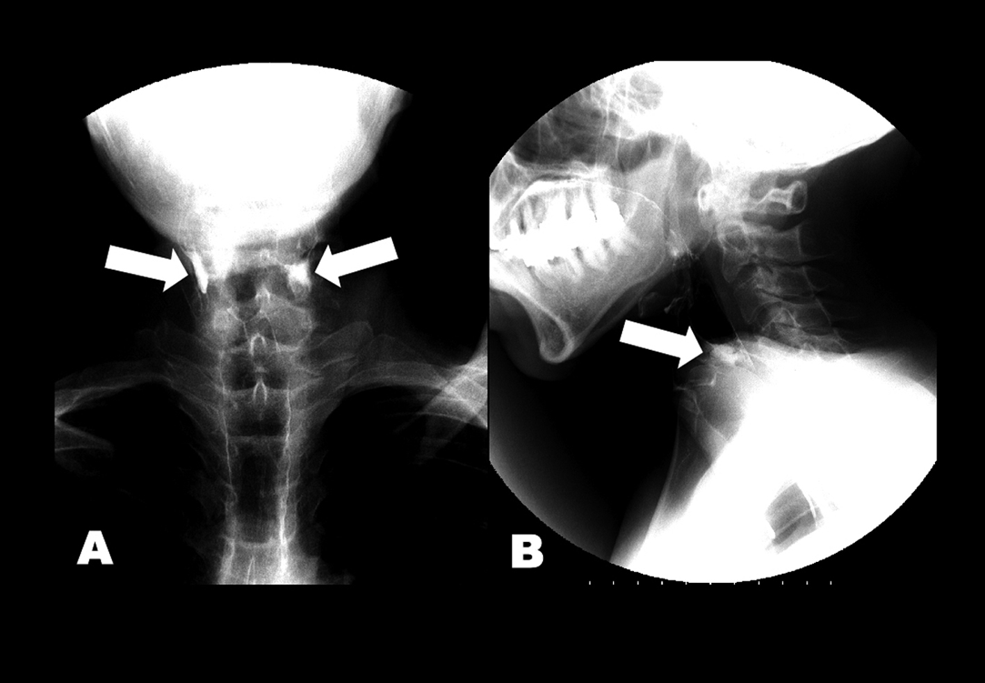
Figure 1. (A) Anteroposterior and (B) lateral views of esophagram. Barium retained at the level of esophageal entrance (arrows) and entered the trachea.
Figure 2.
Figure 2. EMG of the left CPM before and after BTX treatment. (A) Incomplete inhibition of muscle contraction was present during the hypopharyngeal phase of swallowing (between arrows). (B) After BTX injection, CPM contraction amplitudes were decreased at rest compared to pre-treatment of BTX (upper green trace). Normal inhibition of CPM contraction was present during swallowing (between arrowheads).
Figure 3.
Figure 3. Saggital views of spinal cord T2-weighted imaging.Widespread syringomyelia was found from the lumbar to high cervical cord.
Figure 4.
Figure 4. Follow-up T2-weighted imaging before and after onset of dysphagia. (A) No medullary syrinx at 2 years ago. (B) The left medullary syrinx was present at 6 months ago. (C) Axial, (D) sagittal and (E) coronal views of heavy T2-weighted spin-echo inverted imaging showed bilateral extension of medullary syrinx after bulbar onset.