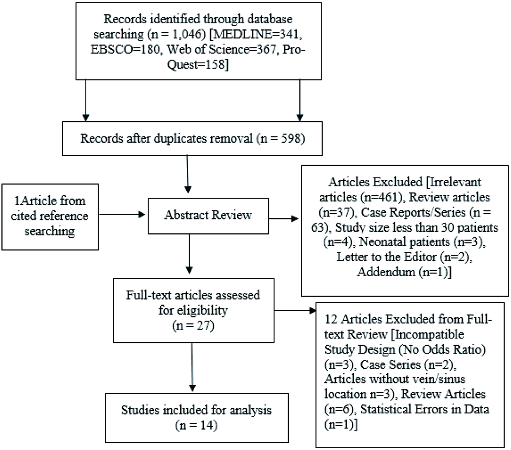
Figure 1. Flow diagram of search process and study selection.
| Journal of Neurology Research, ISSN 1923-2845 print, 1923-2853 online, Open Access |
| Article copyright, the authors; Journal compilation copyright, J Neurol Res and Elmer Press Inc |
| Journal website https://www.neurores.org |
Original Article
Volume 12, Number 3, October 2022, pages 114-120
A Novel Radiographic and Clinical Assessment Scoring Tool for Seizure Risk Stratification in the Acute Phase of Cerebral Venous Sinus Thrombosis: A Systematic Review and Meta-Analysis
Figure
Tables
| OR | 95% CI | |
|---|---|---|
| CI: confidence interval. | ||
| Location | ||
| Frontal lobe | 4.85 | 3.52 - 6.68 |
| Parietal lobe | 2.52 | 1.41 - 4.52 |
| Temporal lobe | 1.58 | 0.60 - 4.14 |
| Occipital lobe | 0.99 | 0.62 - 1.59 |
| Sinus type | ||
| Superficial sagittal sinus | 1.97 | 1.17 - 3.30 |
| Cortical vein | 3.16 | 2.18 - 4.58 |
| Transverse (lateral) sinus | 0.60 | 0.50 - 0.73 |
| Straight sinus | 0.63 | 0.40 - 0.90 |
| Sigmoid sinus | 1.11 | 0.74 -1.66 |
| Deep cerebral vein | 0.39 | 0.07 - 2.07 |
| Venous ischemia type | ||
| Hemorrhagic | 3.85 | 3.20 - 4.64 |
| Non-hemorrhagic | 1.49 | 1.13 - 1.98 |
| Symptom or sign | ||
| Headache | 0.35 | 0.18 - 0.66 |
| Confusion | 2.15 | 1.57 - 2.94 |
| Aphasia | 1.18 | 0.47 - 2.95 |
| Motor deficit | 3.07 | 2.66 - 3.55 |
| Papilledema | 0.36 | 0.22 - 0.58 |
| Sensory disturbance | 1.73 | 0.45- 6.67 |
| Nausea/vomiting | 0.77 | 0.58 - 1.03 |
| Risk factors | Score |
|---|---|
| Radiographic finding (R) | |
| Frontal lobe | 1.5 |
| Parietal lobe | 0.5 |
| Cortical veins | 1 |
| Hemorrhagic venous ischemia | 1.5 |
| Clinical presentation (C) | |
| Motor deficit | 0.5 |
| Confusion | 1 |
| RC score | Estimated seizure risk (%) |
|---|---|
| 0 | 40 |
| 1 | 48 |
| 2 | 52 |
| 3 | 60 |
| 4 | 68 |
| 5 | 80 |
| 6 | 92 |