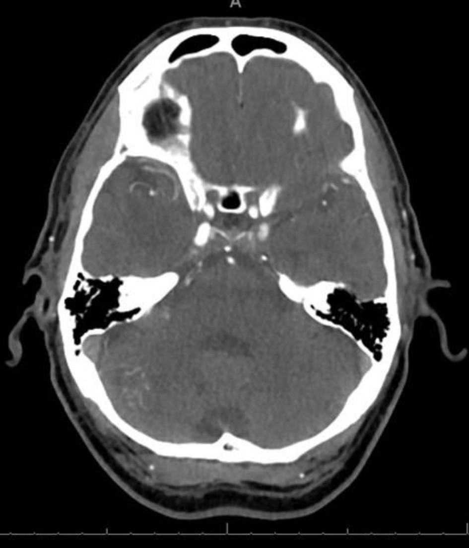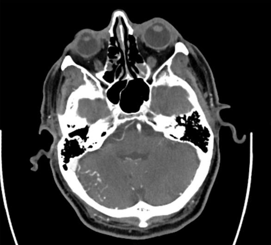
Figure 1. Axial CTA demonstrating serpiginous vessels in the inferolateral aspect of the right cerebellum. CTA: computed tomography angiography.
| Journal of Neurology Research, ISSN 1923-2845 print, 1923-2853 online, Open Access |
| Article copyright, the authors; Journal compilation copyright, J Neurol Res and Elmer Press Inc |
| Journal website https://www.neurores.org |
Case Report
Volume 12, Number 3, October 2022, pages 128-131
Conservative Management for an Infratentorial Pial Arteriovenous Fistula in an Elderly Patient: A Case Report
Figures