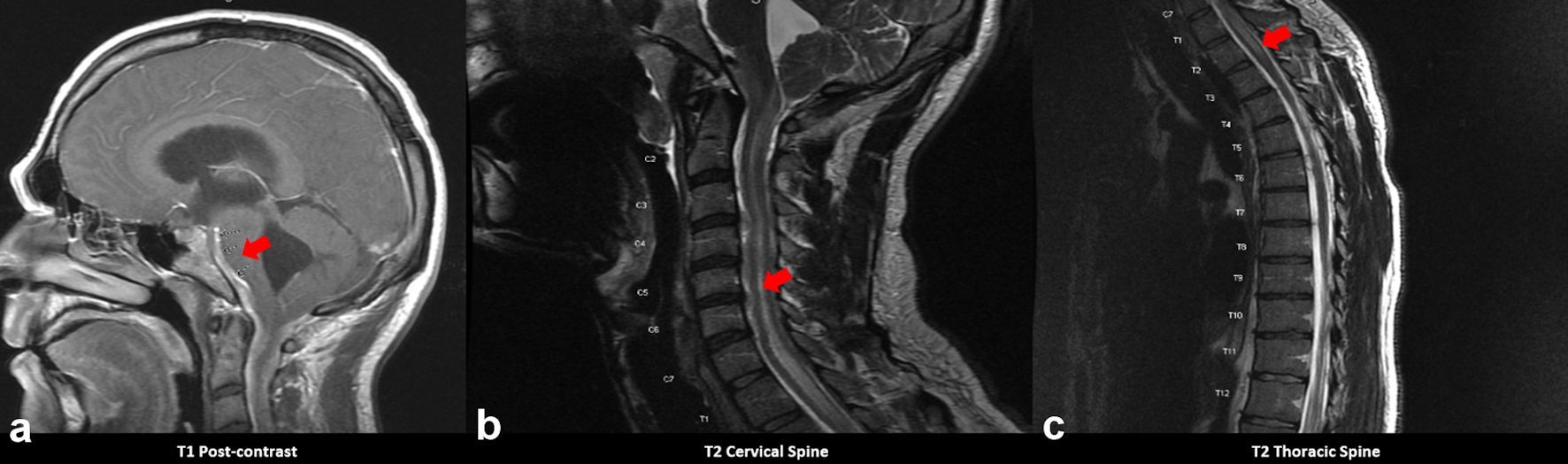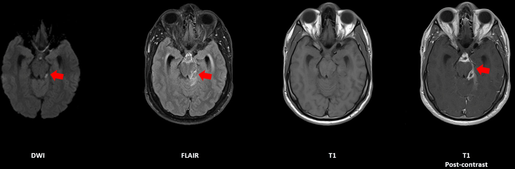
Figure 1. Axial sequences of an MRI brain with and without contrast, demonstrating a rim-enhancing lesion in the suprasellar cistern with restricted diffusion, indicated by red arrows (case 1). MRI: magnetic resonance imaging.
| Journal of Neurology Research, ISSN 1923-2845 print, 1923-2853 online, Open Access |
| Article copyright, the authors; Journal compilation copyright, J Neurol Res and Elmer Press Inc |
| Journal website https://www.neurores.org |
Case Report
Volume 12, Number 1, February 2022, pages 25-29
Diagnostic Dilemma of Aspergillus Meningitis in Patients With Hepatitis C Virus Co-Infection: A Case Series
Figures