Figures
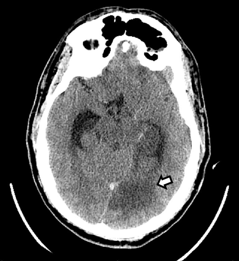
Figure 1. CT of the head without contrast shows an area of hypodensity involving the left medial temporal and occipital lobes, consistent with a subacute ischemic stroke (arrow). CT: computed tomography.
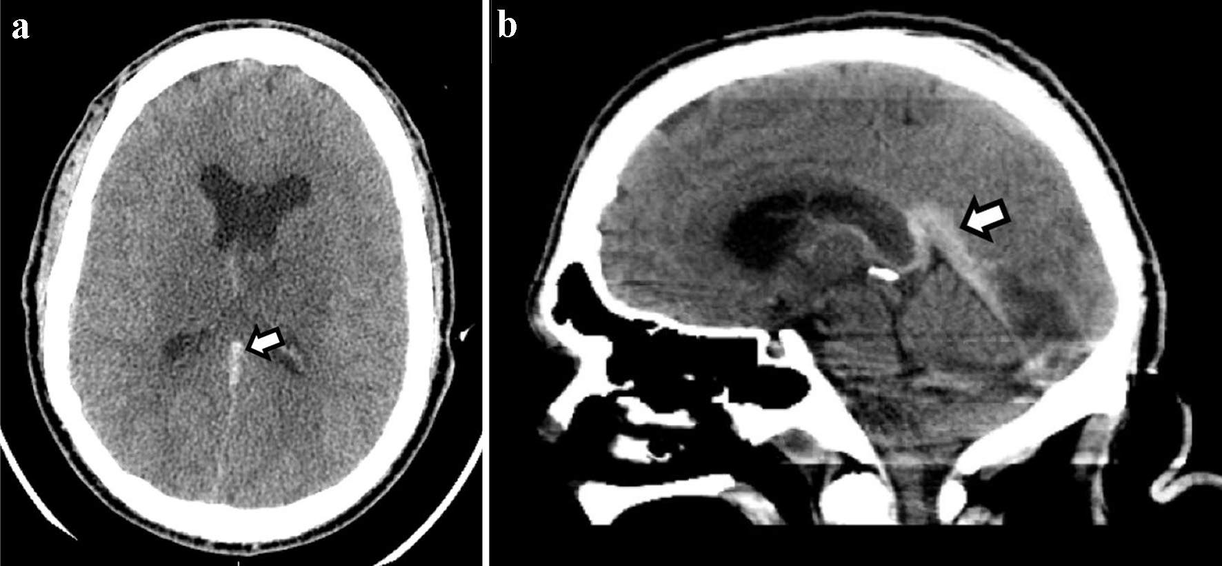
Figure 2. CT of the head without contrast ((a) axial and (b) sagittal)) shows a hyperdense straight sinus consistent with cerebral venous thrombosis. CT: computed tomography.
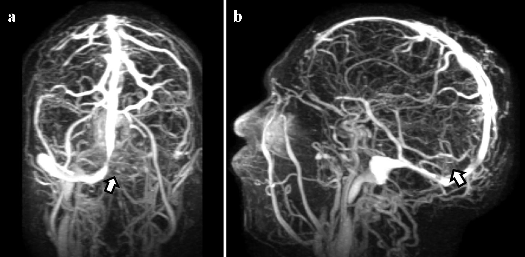
Figure 3. Magnetic resonance venography shows extensive dural venous sinus thrombosis. (a) Presence of occlusive thrombus on the left side involving the transverse, sigmoid, and superior aspect of the internal jugular vein. (b) Thrombosis of the vein of Galen and straight sinus.
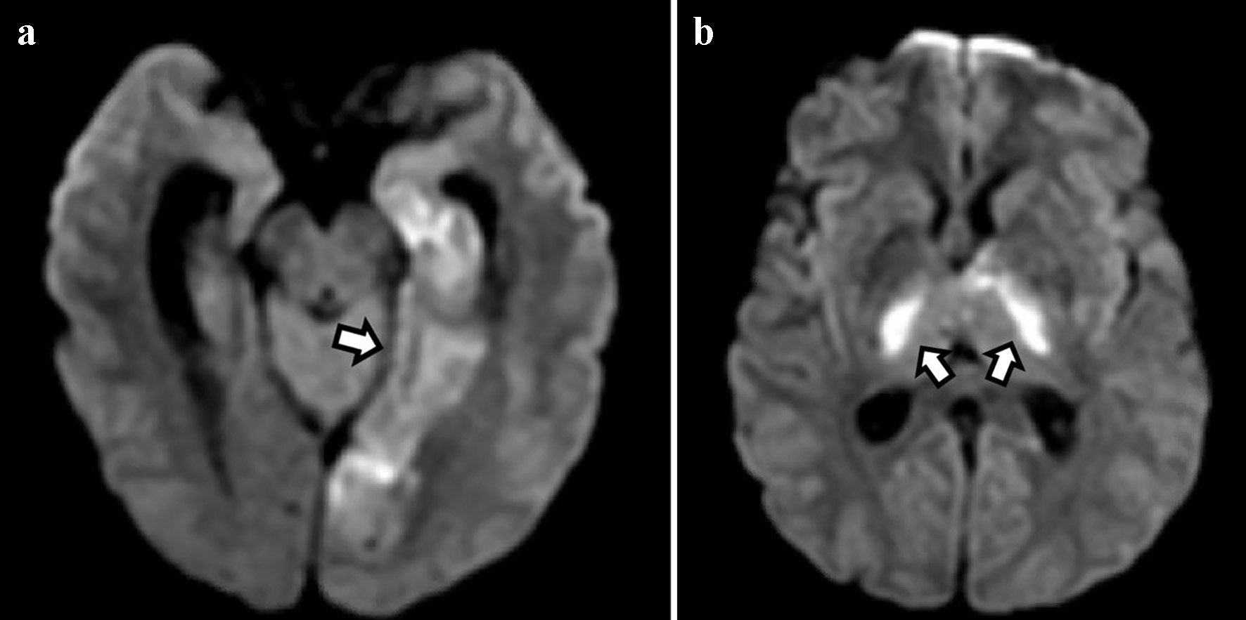
Figure 4. Diffusion-weighted imaging shows areas of restricted diffusion in the medial temporal and occipital lobes (a) and bilateral thalami (b) consistent with acute ischemic stroke.
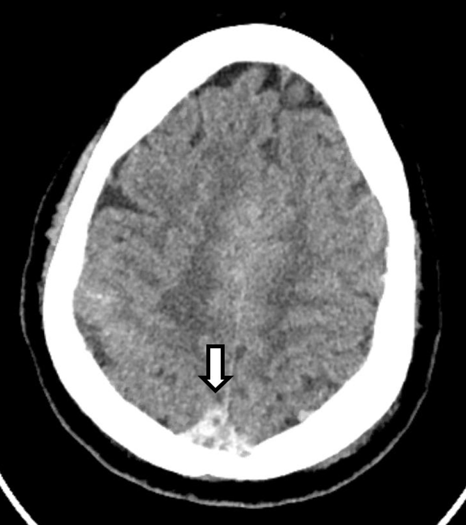
Figure 5. CT of the head without contrast shows an area of hyperdensity involving the superior sagittal sinus (arrow), consistent with a thrombus. CT: computed tomography.
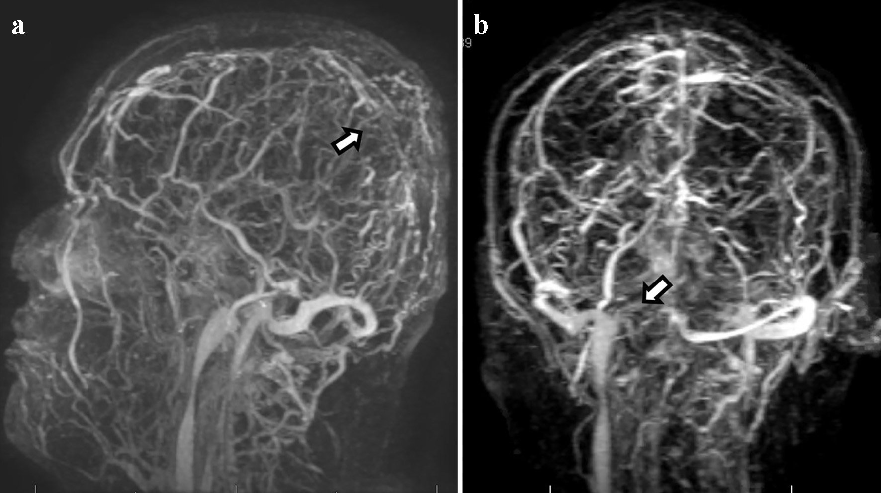
Figure 6. Magnetic resonance venography shows extensive dural venous thrombosis involving multiple transcortical veins along the high convexities, distal two-thirds of the superior sagittal sinus (a), and right-sided transverse and sigmoid sinuses (b).