Journal of Neurology Research, ISSN 1923-2845 print, 1923-2853 online, Open Access
Article copyright, the authors; Journal compilation copyright, J Neurol Res and Elmer Press Inc
Journal website https://www.neurores.org

Original Article

Volume 11, Number 3-4, August 2021, pages 37-46


Patient-Specific Cerebral Flow Model Using Regional Flows and Multi-Objective Optimization

Figures

Figure 1.
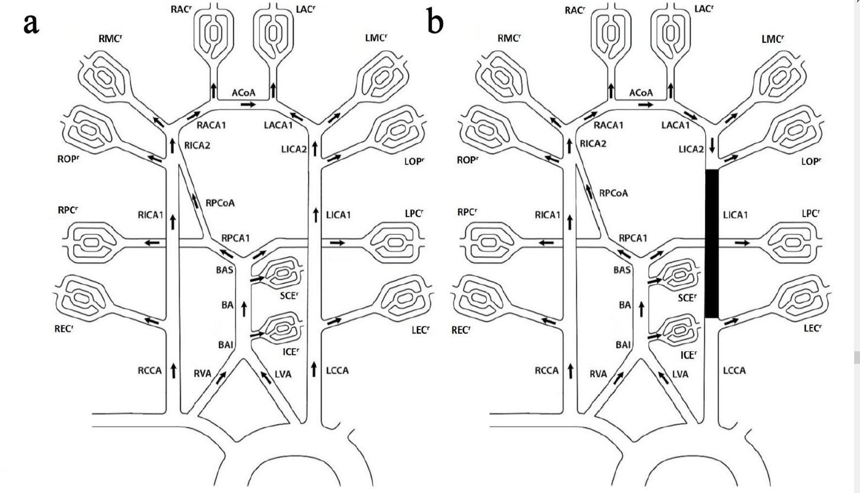
Figure 1. COW without LPCoA. (a) Baseline topology. (b) Left ICA occlusion topology. The 12 regional flows: right and left extra cranial regional flows (RECr and LECr), right and left ophthalmic regional flows (ROPr and LOPr), right and left middle cerebral regional flows (RMCr and LMCr), right and left anterior cerebral arteries section 2 regional flows (RACr and LACr), right and left posterior cerebral artery section 2 regional flows (RPCr and LPCr), anterior inferior cerebella regional flow (ICEr) and superior cerebella regional flow (SCEr); the four influent flows: right and left common carotid arteries (RCCA and LCCA), and right and left vertebral arteries (RVA and LVA); the 12 internal flows: BAI between VA and ICE, basilar artery (BA), BAS between BA and PCA1, right and left internal carotid arteries section 1 and 2 (RICA1, LICA1, RICA2 and LICA2), right posterior artery section 1 (RPCA1), right posterior communicating artery (RPCoA), right and left anterior cerebral arteries section 1 (RACA1 and LACA1), anterior cerebral communicating artery (ACoA). COW: circle of Willis.
Figure 2.
Figure 2. Patient 1’s flow chart and 3D surface rendering. 3D: three-dimensional.
Figure 3.
Figure 3. NOVA flow charts for patient 3 (left) and patient 4 (right).
Figure 4.
Figure 4. Scatter plot comparison between model flows and NOVA flows. The linear fitting R2 = 0.9832. NOVA: Noninvasive Optimal Vessel Analysis.

Tables

Table 1. Vessel Standard Lengths
 
Vessel nameLength (cm)
R/LCCA: right/ left common carotid artery; R/LVA: right/left vertebral artery; BA: basilar artery; R/LICA1: right/left internal carotid artery section 1; R/LPCA1: right/left posterior artery section 1; R/LPCoA: right/left posterior communicating artery; R/LACA1: right/left anterior cerebral artery section 1; ACoA: anterior cerebral communicating artery.
RCCA9.6
LCCA12.1
RVA9.2
LVA7.8
BA3
RPCA10.68
LPCA10.68
RICA117.7
LICA117.7
RPCoA1.35
LPCoA1.35
RACA11.47
LACA11.38
ACoA0.3


Table 2. Patient Demographics/BTO Outcomes/Aortic/Cuff Pressure Measurements
 
PatientGenderAgeAneurysm locationAortic pressure (mm Hg)Cuff pressure (mm Hg)BTO pass/fail
BTO: balloon test occlusion; R/LICA: right/left internal carotid artery.
1Female38LICA10394Passed
2Female78LICA108100Failed
3Male32LICA9989Passed
4Male49RICA9893Failed


Table 3. Patient 1 Diameters and Flows Comparison Between NOVA and Model
 
Vessel nameNOVA diameter (cm)Optimal diameter (cm)NOVA flow (mL/min)Model flow (mL/min)Flow using initial RModel flow error (%)Flow error using intial RInitial R (mm Hg·min/mL)Optimal R (mm Hg·min/mL)
*Using RPCA2’s diameter, no flow measurement. R/LCCA: right/ left common carotid artery; R/LVA: right/left vertebral artery; BA: basilar artery; R/LICA1: right/left internal carotid artery section 1; RPCA1: right posterior artery section 1; RPCoA: right posterior communicating artery; R/LACA1: right/left anterior cerebral artery section 1; ACoA: anterior cerebral communicating artery.
RCCA0.590.6067339325312-4-80.001990.00178
LCCA0.530.532312298301-5-40.002430.00239
RVA0.370.3476100100106060.00980.01258
LVA0.320.3007666670060.014850.01905
BA0.330.307139139149080.005050.00674
RPCA10.2*0.1887-4858--0.008490.01071
RICA10.460.4893261247234-5-100.007890.00617
LICA10.490.5052299285288-5-40.006130.00543
RPCoA0.130.1349-8-8201250.094370.08147
RACA10.270.2623897572-16-190.005520.0062
LACA10.250.2466756164-19-150.007050.00745
ACoA0.130.1276630-500.020970.02302


Table 4. Peripheral Regional Resistance at Baseline for Patient 1
 
RegionResults (mm Hg·min/mL)
R/LEC: right/left external carotid artery; R/LPC: right/left posterior cerebral artery; R/LMC: right/left middle cerebral artery; R/LAC: right/left anterior artery; SCE: superior cerebellar artery; ICE: inferior cerebellar artery; R/LOP: right and left ophthalmic artery.
REC1.18
RPC1.61
ROP9.09
RMC0.59
RAC1.31
LAC1.35
LMC0.68
LOP1.0
LPC1.33
SCE3.95
ICE3.4
LEC7.1


Table 5. SPs Comparison Between Model and Clinical Measurement
 
PatientClinical (mm Hg)Model (mm Hg) with aortic pressureChange (%)Model (mm Hg) with cuff pressureChange (%)
SP: stump pressure.
19588-781-15
27062-1157-19
39295385-8
469747701


Table 6. Flow and Pressure Comparison Between Baseline and Occlusion Model for Patient 1
 
Region/vesselFlow (mL/min)Pressure (mm Hg)
BaselineOcclusionChange (%)BaselineOcclusionChange (%)
RECr: right extra cranial regional flow; ROPr and LOPr: right and left ophthalmic regional flows; RMCr and LMCr: right and left middle cerebral regional flows; RACr and LACr: right and left anterior cerebral arteries regional flows; RPCr and LPCr: right and left posterior cerebral artery regional flows; ICEr: anterior inferior cerebella regional flow; SCEr: superior cerebella regional flow; RCCA: right common carotid artery; RVA and LVA: right and left vertebral arteries; BA: basilar artery; RICA1: right internal carotid artery section 1; RPCA1: right posterior artery section 1; RPCoA: right posterior communicating artery; RACA1 and LACA1: right and left anterior cerebral arteries section 1; ACoA: anterior cerebral communicating artery.
RECr787801021020
RPCr565601001000
ROPr1010010199-2
RMCr154151-210199-2
RACr6966-410096-4
LACr6760-1010090-10
LMCr133117-1210188-13
LOPr9180-1210188-13
LPCr68680101100-1
SCEr23230101100-1
ICEr272701021020
RCCA325552701031030
RVA100110101021020
LVA6673111021020
BA139156121011010
RPCA1486535101100-1
RICA12474759210298-4
RPCoA-8921310199-2
RACA17532333110198-3
LACA16119722310190-11
ACoA62574,18310094-6