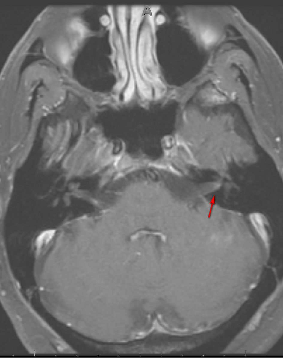
Figure 1. Axial MRI of the brain, T1 with gadolinium showing enhancement of CN VII on the right (arrow). MRI: magnetic resonance imaging; CN: cranial nerve.
| Journal of Neurology Research, ISSN 1923-2845 print, 1923-2853 online, Open Access |
| Article copyright, the authors; Journal compilation copyright, J Neurol Res and Elmer Press Inc |
| Journal website https://www.neurores.org |
Case Report
Volume 11, Number 1-2, April 2021, pages 27-31
An Early Presentation of Neurosyphilis Manifesting as Cranial Polyneuropathies: A Case Report
Figures