Journal of Neurology Research, ISSN 1923-2845 print, 1923-2853 online, Open Access
Article copyright, the authors; Journal compilation copyright, J Neurol Res and Elmer Press Inc
Journal website https://www.neurores.org

Case Report

Volume 10, Number 6, December 2020, pages 248-252


Sudden Onset Hemiplegia and Neglect: A Case Report of Type A Aortic Dissection Presenting as a Code Stroke

Figures

Figure 1.
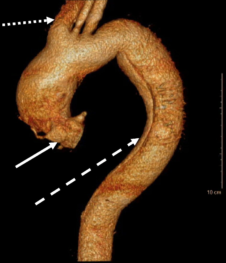
Figure 1. CT angiogram thoracic aorta three-dimensional (3D) reconstruction demonstrating Stanford type A AAD at the aortic root (solid white arrow), with extension into the innominate artery (dotted white arrow) and distal extension to the descending thoracic aorta (dashed white arrow). CT: computed tomography.
Figure 2.
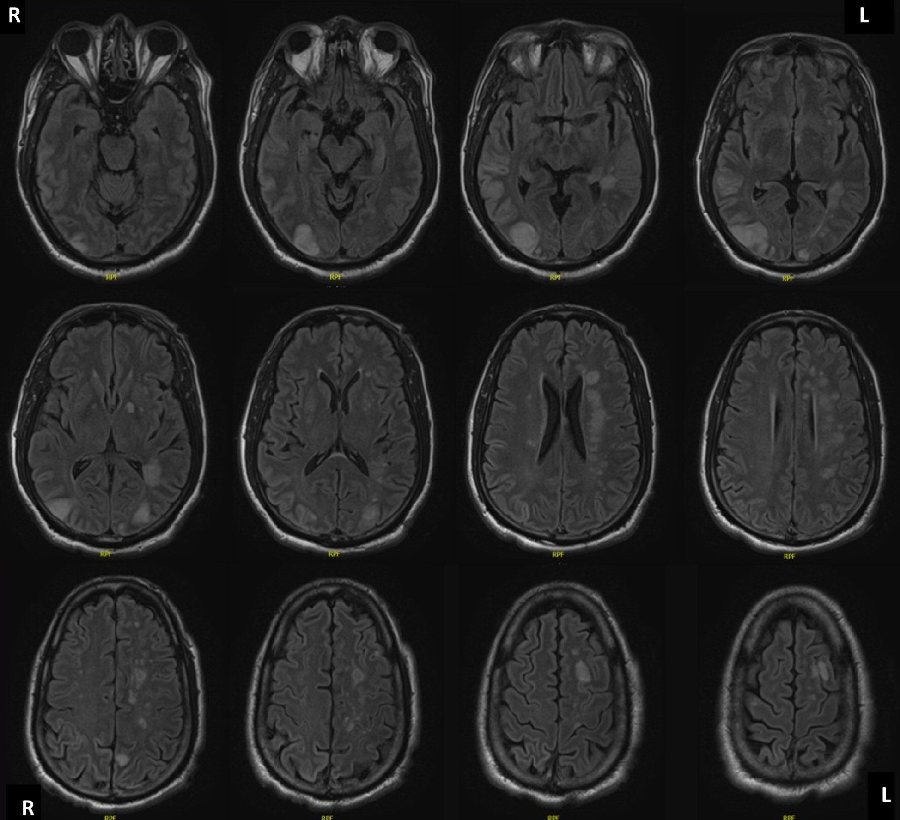
Figure 2. MRI brain without contrast, axial T2 weighted imaging, demonstrating diffuse regions of increased relaxation time in the left periventricular white matter, left frontal, right temporal, left cerebellar hemisphere, left parietal, and bilateral occipital lobes, and left hypothalamus, consistent with global watershed ischemia and infarction territories. MRI: magnetic resonance imaging.

Table

Table 1. Presenting Initial NIH Stroke Scale (NIHSS) Score as Determined by Consulting Neurology Stroke Service.
 
This patient’s NIHSS is 17, which is categorized as severe. NIH: National Institutes of Health.
Level of consciousness0
Level of consciousness questions0
Level of consciousness commands0
Extraocular movements2
Visual fields2
Facial palsy0
Left arm motor drift4
Right arm motor drift0
Left leg motor drift4
Right leg motor drift0
Limb ataxia2
Sensation0
Language/aphasia0
Dysarthria1
Extinction/inattention2
Total17