Journal of Neurology Research, ISSN 1923-2845 print, 1923-2853 online, Open Access
Article copyright, the authors; Journal compilation copyright, J Neurol Res and Elmer Press Inc
Journal website https://www.neurores.org

Review

Volume 10, Number 5, October 2020, pages 173-176


Approach to Brain Magnetic Resonance Imaging for Non-Radiologists

Figures

Figure 1.
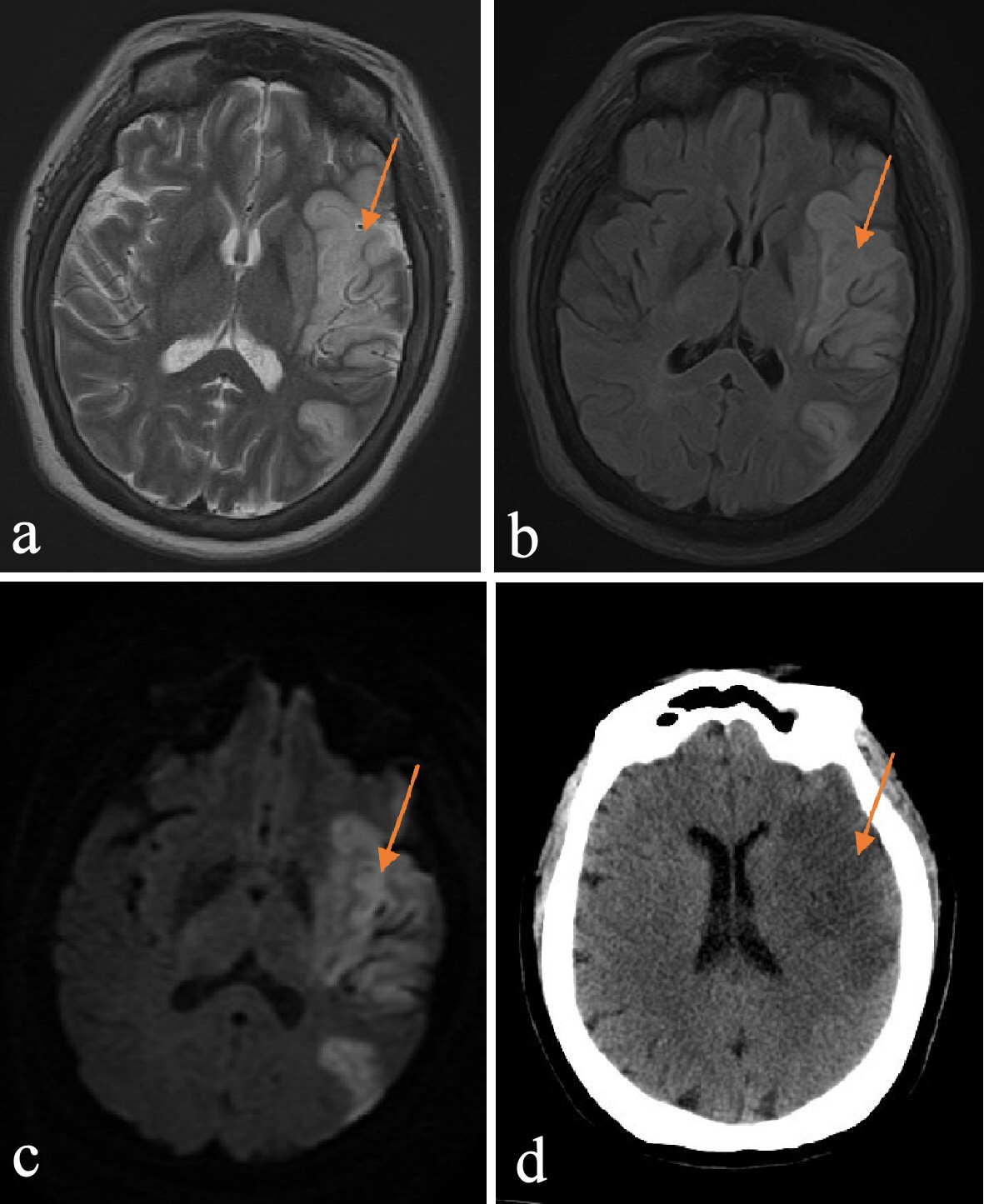
Figure 1. (a) T2 image of left MCA territory infarct, with associated vasogenic edema. (b) FLAIR image of left MCA territory infarct, with associated vasogenic edema. (c) DWI image of left MCA territory infarct, with associated vasogenic edema. (d) CT image of left MCA territory infarct, with associated vasogenic edema. MCA: middle cerebral artery; FLAIR: fluid attenuation inversion recovery; DWI: diffusion-weighted imaging; CT: computed tomography.
Figure 2.
Figure 2. (a) DWI image of ischemic stroke with hemorrhagic transformation. (b) SWI image of ischemic stroke with hemorrhagic transformation. (c) T2 image of ischemic stroke with hemorrhagic transformation. DWI: diffusion-weighted imaging; SWI: susceptibility-weighted imaging.
Figure 3.
Figure 3. CT image of acute infarct in cerebellum and occipital lobe. CT: computed tomography.