Figures
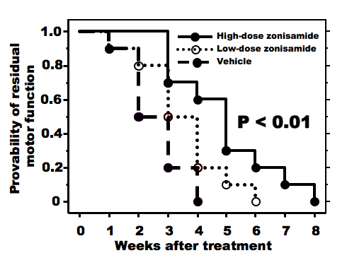
Figure 1. Progression of forelimb motor deficits. High-dose zonisamide administration retards progression of forelimb dysfunction significantly. (P < 0.01 by Kaplan-Meyer method).
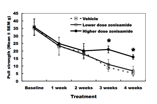
Figure 2. Pull-strength of the forelimbs. High-dose zonisamide administration delays decline of pull-strength at 3 and 4 weeks after treatment. (*P < 0.01 at 3 and 4 weeks after treatment by unpaired Student’s t-test).
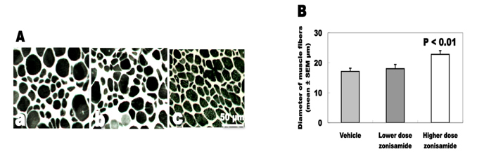
Figure 3. Denervation changes of the biceps muscles. (A) ATPase staining of biceps muscles. (a) Vehicle treatment. (b) Low-dose zonisamide treatment. (c) High-dose zonisamide treatment. (B) Treatment with high-dose zonisamide increases the mean diameter of muscle fibers at 30%. (P < 0.01 between the high-dose zonisamide group and the vehicle group by one-way ANOVA followed by Scheffe’s test)
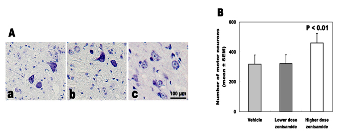
Figure 4. Total number of survived motor neurons. (A) Nissl staining of C5-6 cord. (a) Vehicle treatment. (b) Low-dose zonisamide treatment. (c) High-dose zonisamide treatment. (B) High-dose zonisamide administration inhibits the loss of motor neurons at 45%. (P < 0.01 between the high-dose zonisamide group and the vehicle group by one-way ANOVA followed by Scheffe’s test)
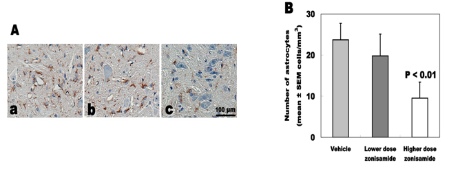
Figure 5. Number of GFAP-immunoreactive astrocytes. (A) GFAP immunostaining of C5-6 cord. (a) Vehicle treatment. (b) Low-dose zonisamide treatment. (c) High-dose zonisamide treatment. (B) High-dose zonisamide treatment decreases degree of astrocyte proliferation at 60%. (P < 0.01 between the high-dose zonisamide and the vehicle group by one-way ANOVA followed by Scheffe’s test)