Journal of Neurology Research, ISSN 1923-2845 print, 1923-2853 online, Open Access
Article copyright, the authors; Journal compilation copyright, J Neurol Res and Elmer Press Inc
Journal website http://www.neurores.org

Case Report

Volume 10, Number 4, August 2020, pages 136-139


Endovascular Reconstruction Utilizing Flow Diversion Stenting in a Patient With Bilateral Giant Cavernous Internal Carotid Artery Aneurysms

Figures

Figure 1.
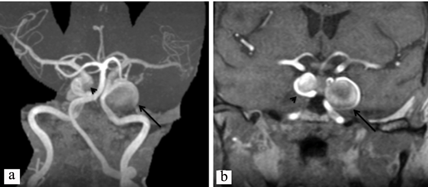
Figure 1. (a) Magnetic resonance angiography of the head showing a giant aneurysm of the left ICA (arrow) and a smaller aneurysm in the cavernous ICA on the right (arrow head). (b) Magnetic resonance imaging, T1, with gadolinium, showing a giant cavernous ICA aneurysm on the left (black arrow) and a smaller aneurysm in the cavernous ICA on the right (arrow head). ICA: internal carotid artery.
Figure 2.
Figure 2. (a) Cerebral angiogram showing a giant right ICA aneurysm before treatment (black arrow). (b, c) Post-treatment with successful flow diversion (black arrow in (c)). (d-f) Six months post treatment, stable imaging and a left carotid stent (white arrow in (f)). ICA: internal carotid artery.