Journal of Neurology Research, ISSN 1923-2845 print, 1923-2853 online, Open Access
Article copyright, the authors; Journal compilation copyright, J Neurol Res and Elmer Press Inc
Journal website http://www.neurores.org

Case Report

Volume 10, Number 3, June 2020, pages 99-103


Use of Continuous Infusions of Eptifibatide and Cangrelor in Large Vessel Occlusion Acute Ischemic Stroke After Emergent Carotid Artery Stenting

Figures

Figure 1.
Figure 1. Diagnostic cerebral angiogram (AP view) demonstrating critical left ICA stenosis associated with the dissection in the ascending cervical segment of the left ICA prior to balloon angioplasty and stenting (arrow). ICA: internal carotid artery; AP: anteroposterior.
Figure 2.
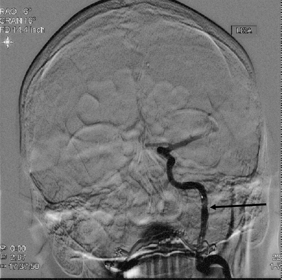
Figure 2. Diagnostic cerebral angiogram (AP view) following balloon angioplasty and stent placement in the ascending cervical segment of the left ICA, showing significantly improved contrast opacification of the artery (arrow). ICA: internal carotid artery; AP: anteroposterior.
Figure 3.
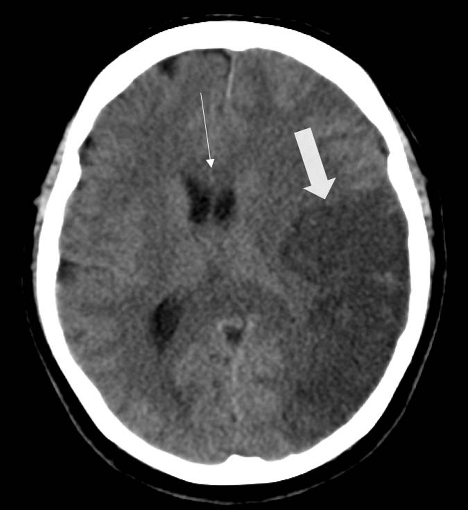
Figure 3. Head CT (axial view) exhibiting hypodensity within the left temporal lobe of the brain, consistent with the left M2 inferior division occlusion (thick arrow). The scan, performed less than 36 h out from the revascularization procedure, already demonstrates subtle midline shift and mass effect from the area of infarcted brain (thin arrow). CT: computed tomography.