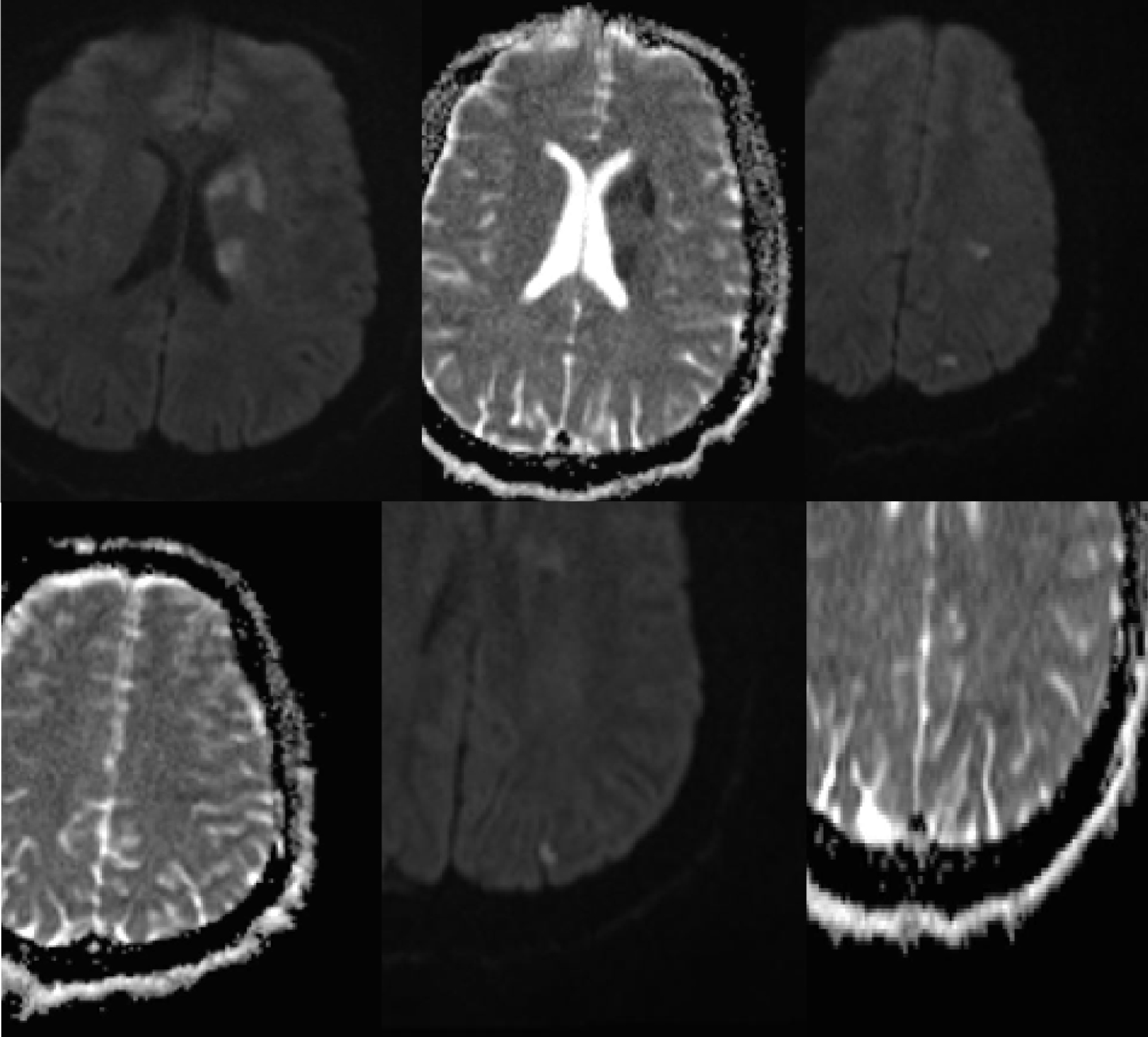
Figure 1. Multiple foci of diffusion restriction in the left hemisphere which are mostly in the basal ganglia and scattered in white matter of the frontal, parietal and occipital lobes.
| Journal of Neurology Research, ISSN 1923-2845 print, 1923-2853 online, Open Access |
| Article copyright, the authors; Journal compilation copyright, J Neurol Res and Elmer Press Inc |
| Journal website http://www.neurores.org |
Case Report
Volume 10, Number 3, June 2020, pages 95-98
Unilateral Cardioembolic Stroke in a Patient With Aortic Arch Anomaly
Figures