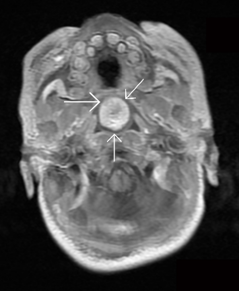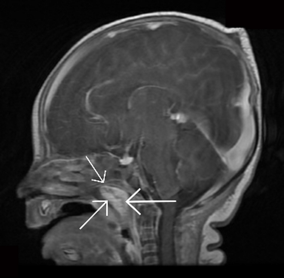
Figure 1. Brain MRI (sagittal view): the arrows show the mass in the nasopharyngeal area occluding the airway.
| Journal of Neurology Research, ISSN 1923-2845 print, 1923-2853 online, Open Access |
| Article copyright, the authors; Journal compilation copyright, J Neurol Res and Elmer Press Inc |
| Journal website http://www.neurores.org |
Case Report
Volume 10, Number 2, April 2020, pages 48-51
Nasopharyngeal Teratoma in a Neonate
Figures