Journal of Neurology Research, ISSN 1923-2845 print, 1923-2853 online, Open Access
Article copyright, the authors; Journal compilation copyright, J Neurol Res and Elmer Press Inc
Journal website http://www.neurores.org

Review

Volume 10, Number 2, April 2020, pages 32-37


Electroencephalography During the Acute Phase Of Encephalitis: A Brief Review

Figures

Figure 1.
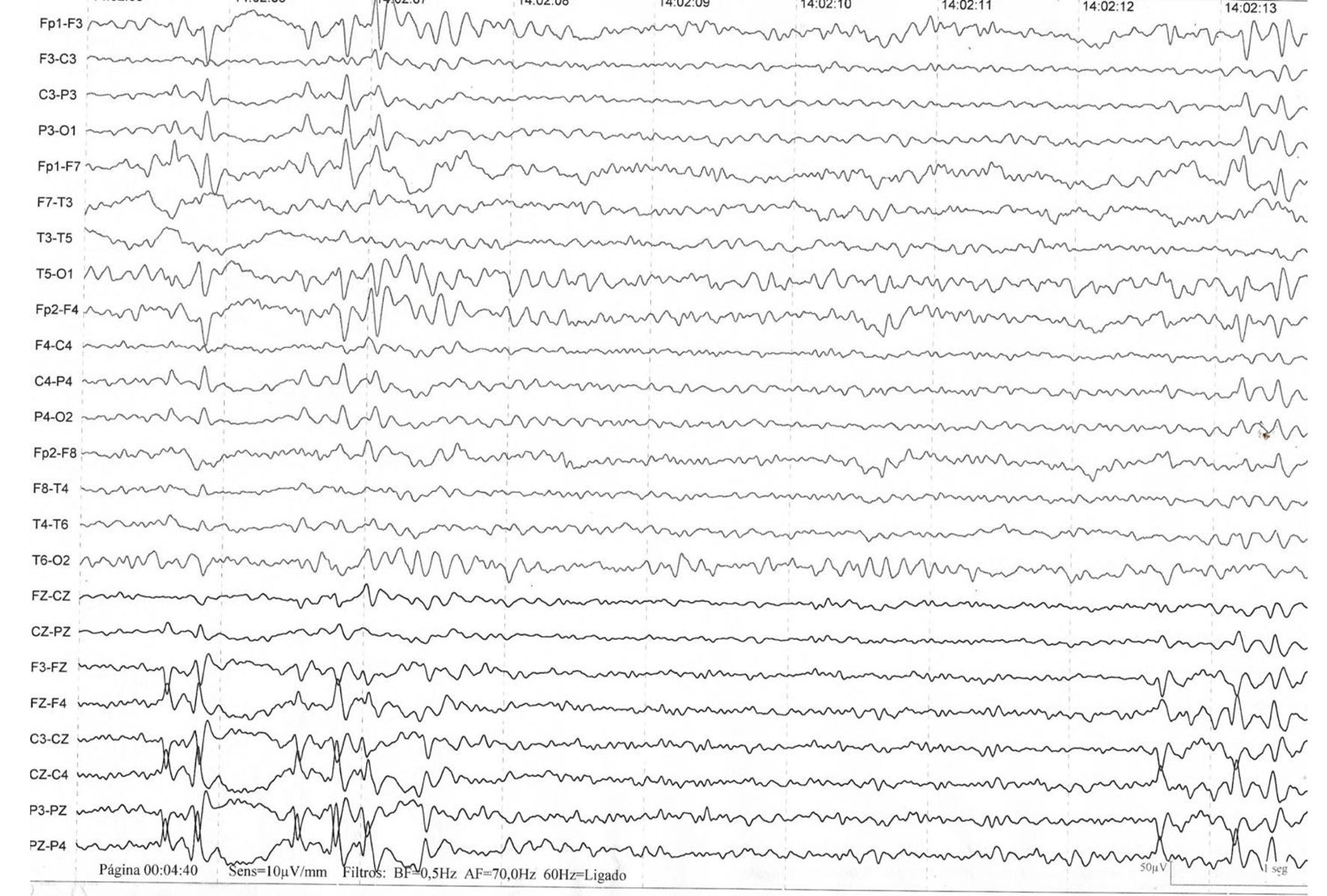
Figure 1. The presence of three-phase waves are common but nonspecific. They occur in multiple encephalopathies and metabolic alterations of various conditions, such as hepatic, uremic and septic encephalopathies, but may also be present during the course of viral encephalitis or another etiology [1, 9].
Figure 2.
Figure 2. The presence of bilateral slow thriphasic waves in a patient affected by West Nile encephalitis. This trace is similar to referenced in literature [10].
Figure 3.
Figure 3. Generalized activity of rhythmic and semi-rhythmic delta frequency at 1 Hz with superimposed bursts, predominantly frontal of rhythmic beta frequency activity.
Figure 4.
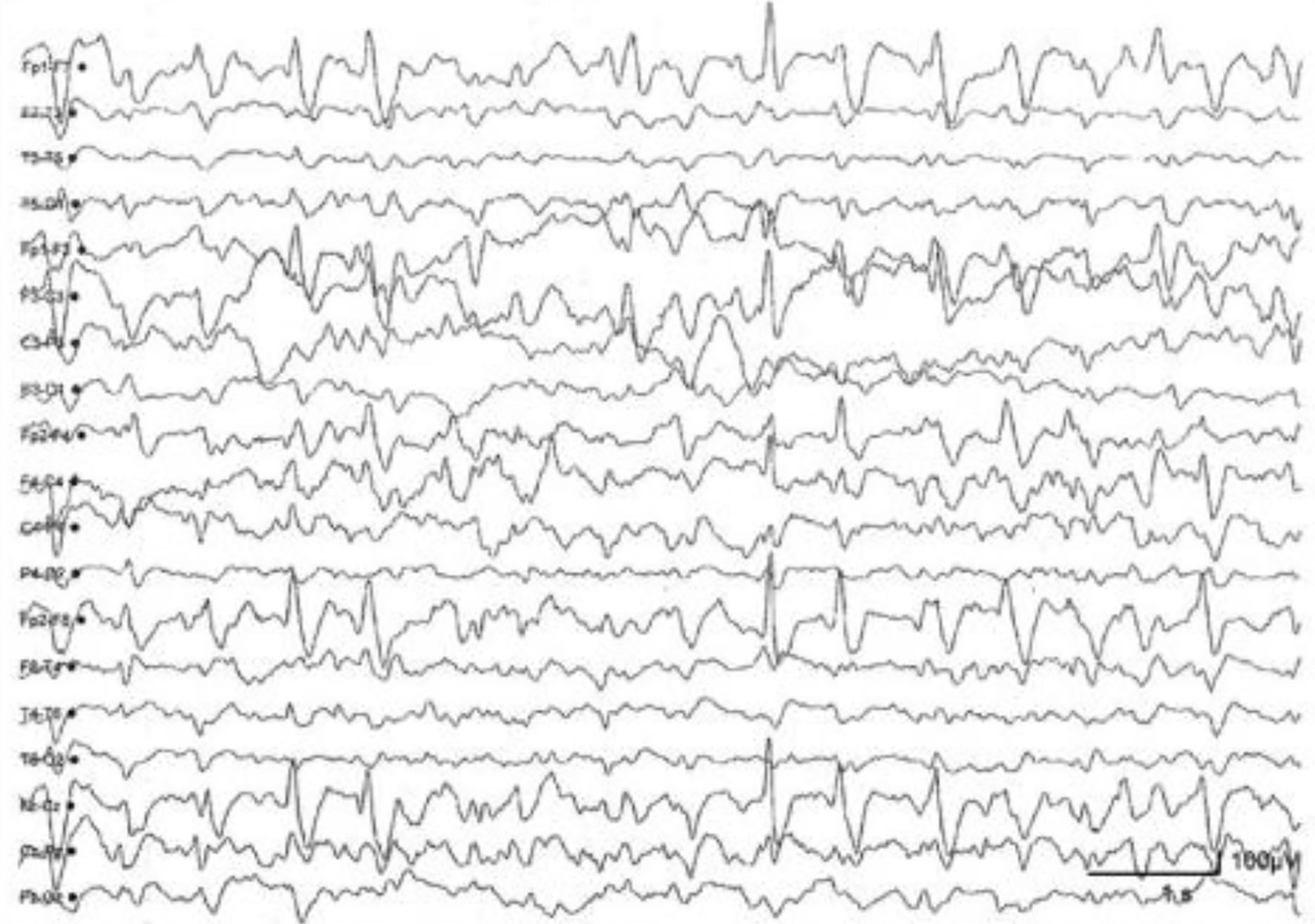
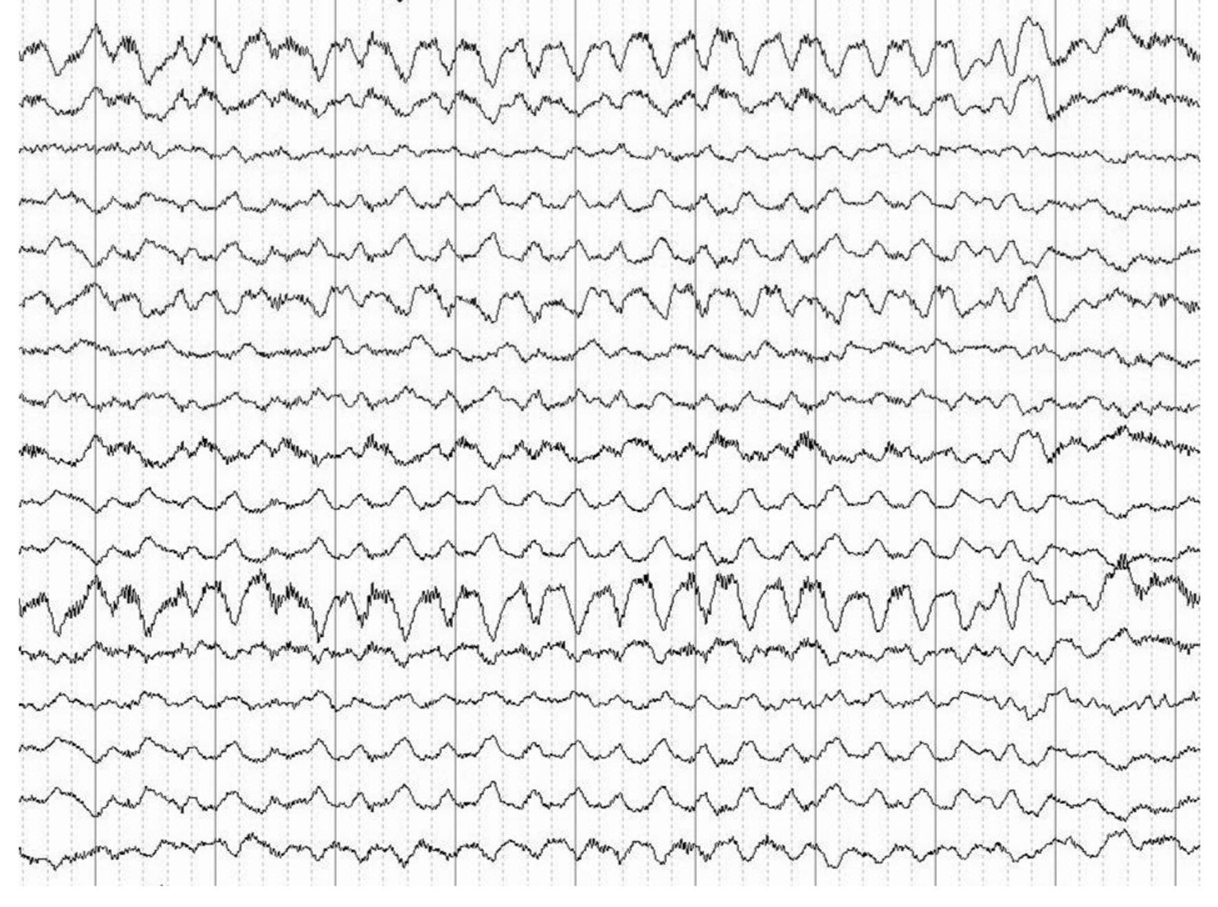
Figure 4. Anti-NMDA receptor encephalitis. Generalized irregular activity of 1.5 to 2 Hz peak and predominant wave in the previous regions of the hemisphere and slow and diffuse background activity. The record shows that the extreme delta brush pattern is not pathognomonic or unique sign of EEG in anti-NMDA receptor encephalitis. NMDA: N-methyl-D-aspartate; EEG: electroencephalography.
Figure 5.
Figure 5. (a) qEEG monitoring showing the electrical activity in status epilepticus due to traumatic brain injury. (b) EEG showing no electroencephalographic seizure. (c) EEG showing diffuse electroencephalographic seizures. qEEG: quantitative electroencephalography.