Journal of Neurology Research, ISSN 1923-2845 print, 1923-2853 online, Open Access
Article copyright, the authors; Journal compilation copyright, J Neurol Res and Elmer Press Inc
Journal website http://www.neurores.org

Case Report

Volume 10, Number 1, February 2020, pages 17-19


Dramatic Recovery of a Patient With Wernicke Encephalopathy Presenting With the Lesion of the Corpus Callosum

Figure

Figure 1.
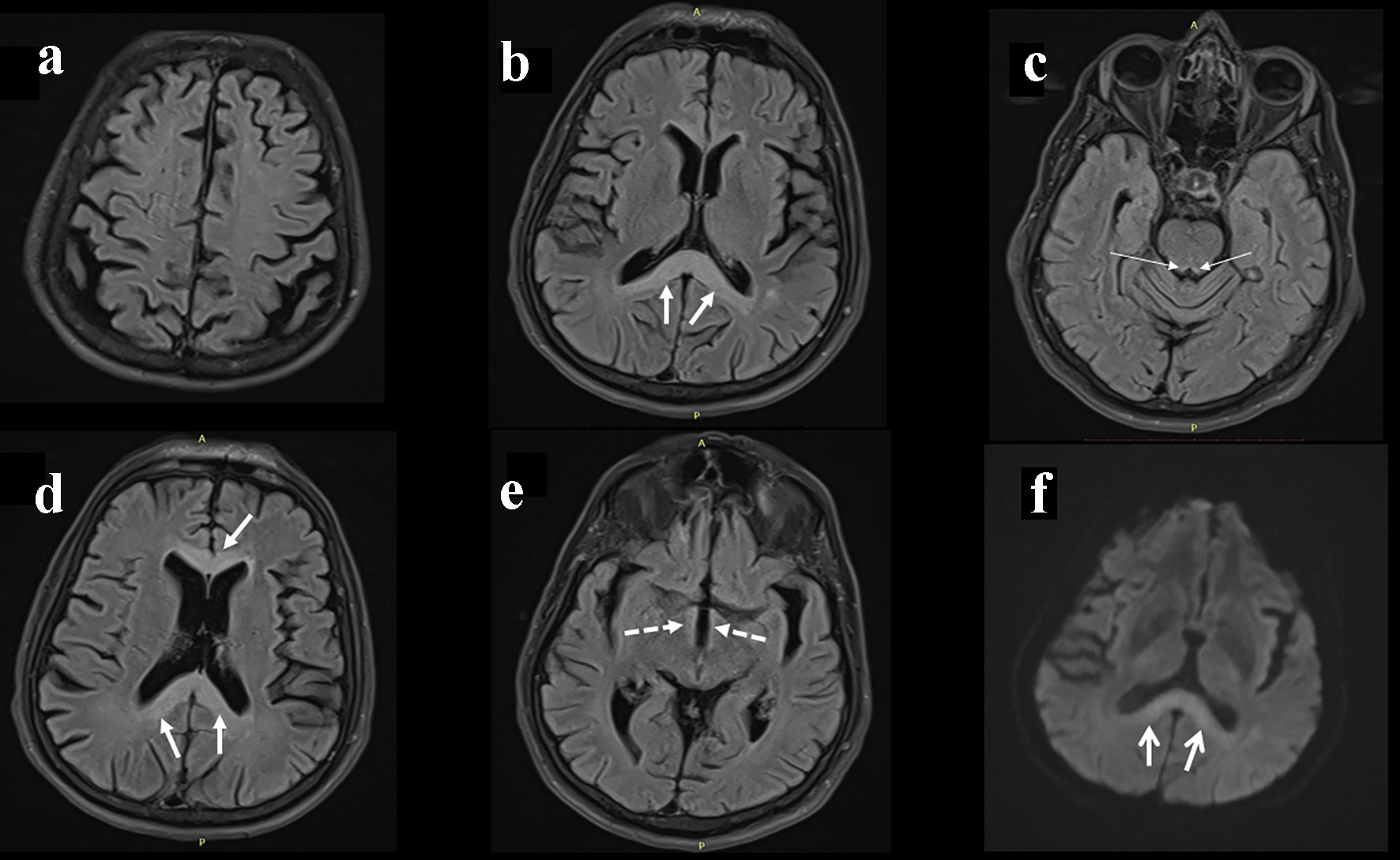
Figure 1. Cranial magnetic resonance image shows T2 hyperintense lesions in the genu and splenium of the corpus callosum (b, d), mamillar body (e). A slight hyperintensity in the periaqueductal region was also recognized (c). Diffusion-weighted imaging shows high signal in the splenial lesion of the corpus callosum (f) (arrows).