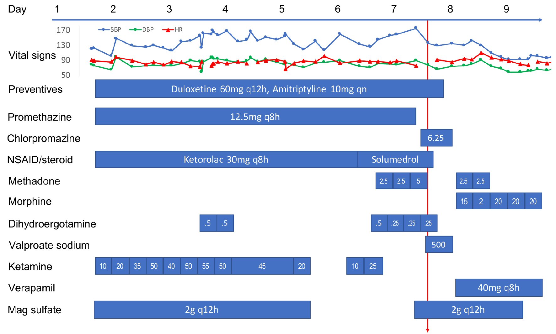
Figure 1. Time course of vital signs and medications administered to the patient during the hospitalization. All medications were intravenous formulation except oral preventives, methadone/morphine, and verapamil. All doses are in mg except where noted. Red arrow indicates the time of PRES/RCVS onset. PRES: posterior reversible encephalopathy syndrome; RCVS: reversible cerebral vasoconstriction syndrome; NSAID: non-steroidal anti-inflammatory drug.
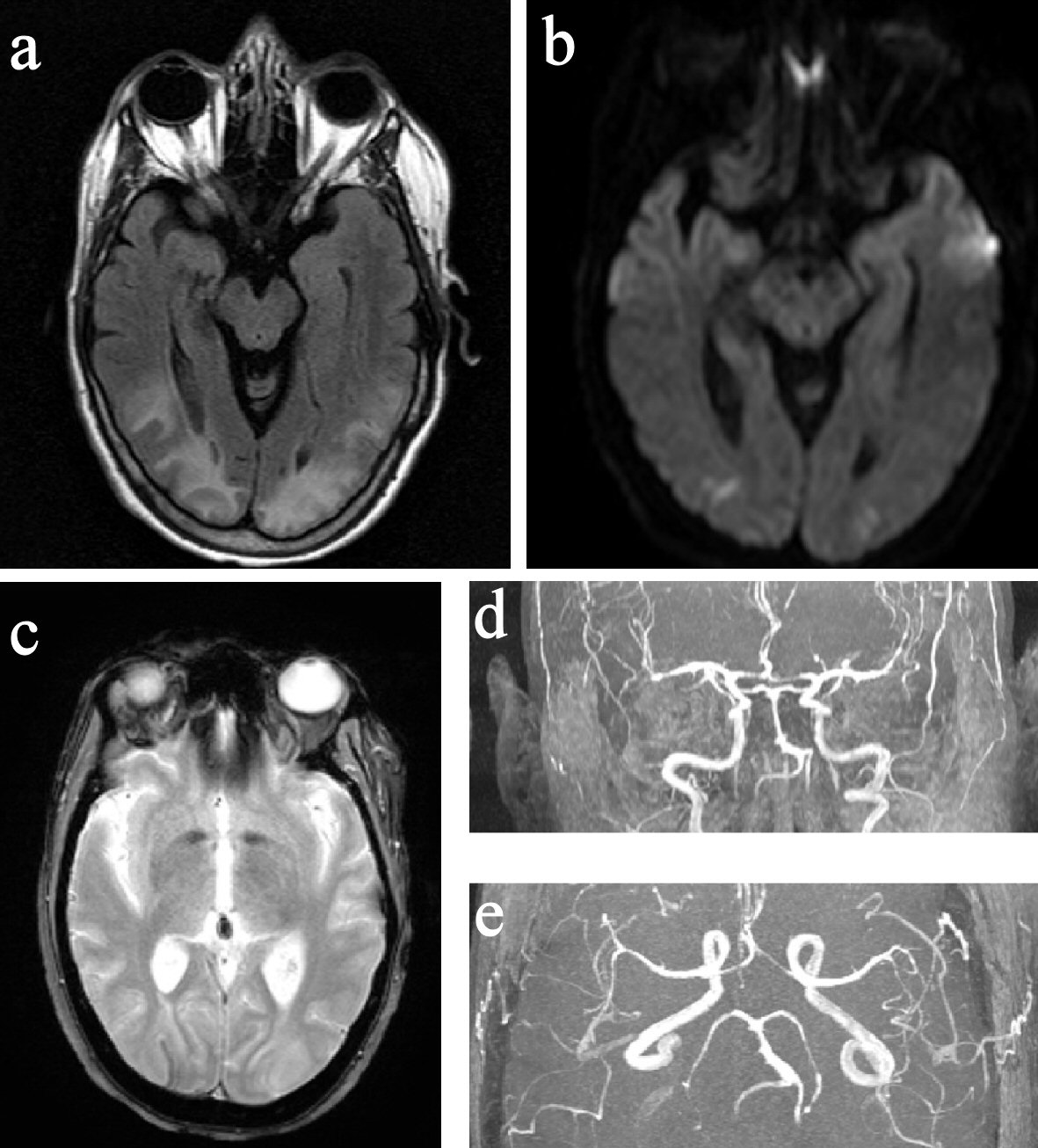
Figure 2. MRI and MRA of the brain. (a) FLAIR image showed hyperintensity in the posterior parietal and occipital lobes. (b) DWI image showed small bilateral occipital pole diffusion restrictions. (c) GRE image showed right occipital curvilinear superficial siderosis. (d) and (e) Segmental narrowing of bilateral ACA, MCA, PCA, and BA. MRI: magnetic resonance imaging; MRA: magnetic resonance angiography; FLAIR: fluid-attenuated inversion recovery; DWI: diffusion-weighted imaging; GRE: gradient-recalled echo; ACA: anterior cerebral artery; MCA: middle cerebral artery; PCA: posterior cerebral artery; BA: basilar artery.