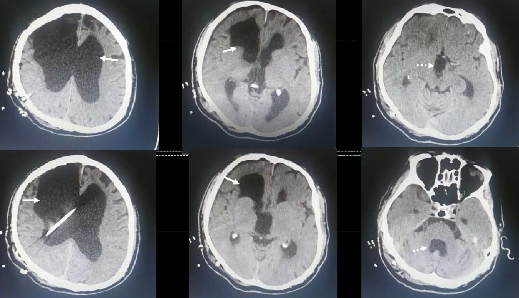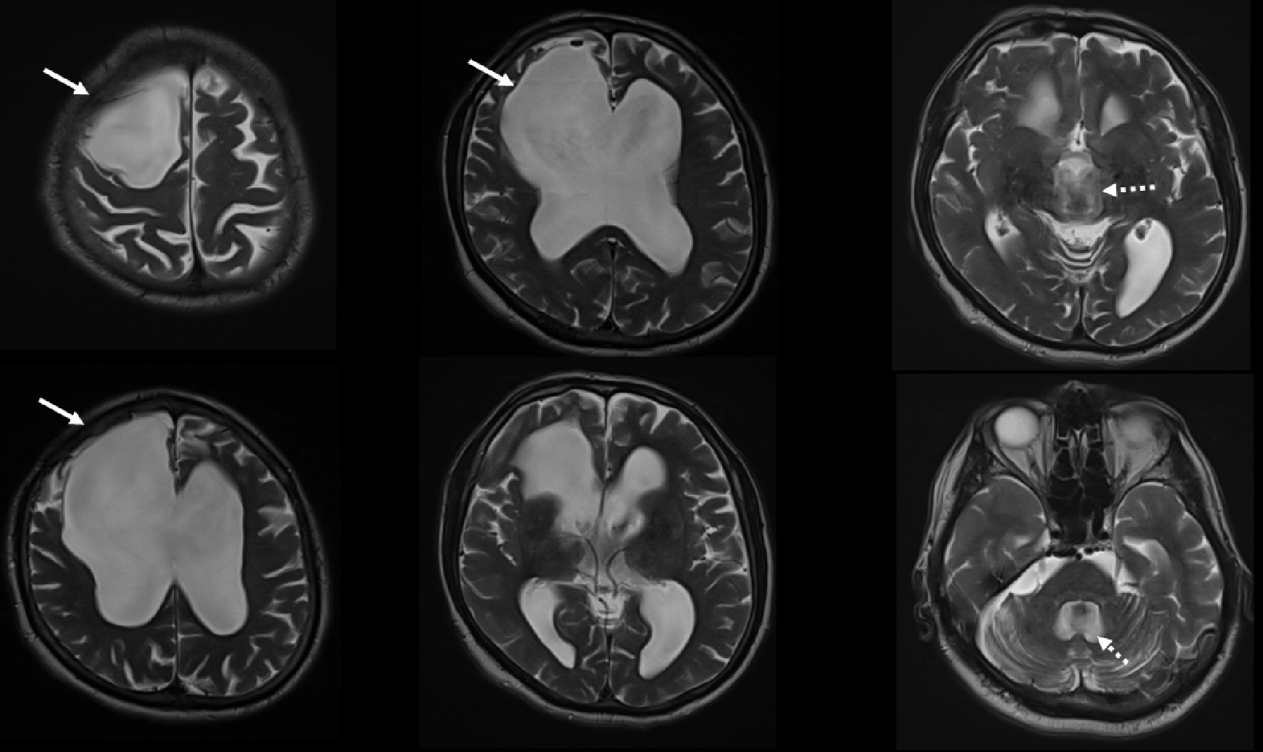
Figure 1. Cranial MRI shows obstructive hydrocephalus and righ frontal encephalomalacia (jagged arrows shows enlargement of third and fourth ventricles). MRI: magnetic resonance imaging.
| Journal of Neurology Research, ISSN 1923-2845 print, 1923-2853 online, Open Access |
| Article copyright, the authors; Journal compilation copyright, J Neurol Res and Elmer Press Inc |
| Journal website http://www.neurores.org |
Letter to the Editor
Volume 9, Number 4-5, October 2019, pages 89-91
A Dramatic Improvement by Ventriculoperitoneal Shunt Surgery in a Patient With Secondary Normal Pressure Hydrocephalus
Figures