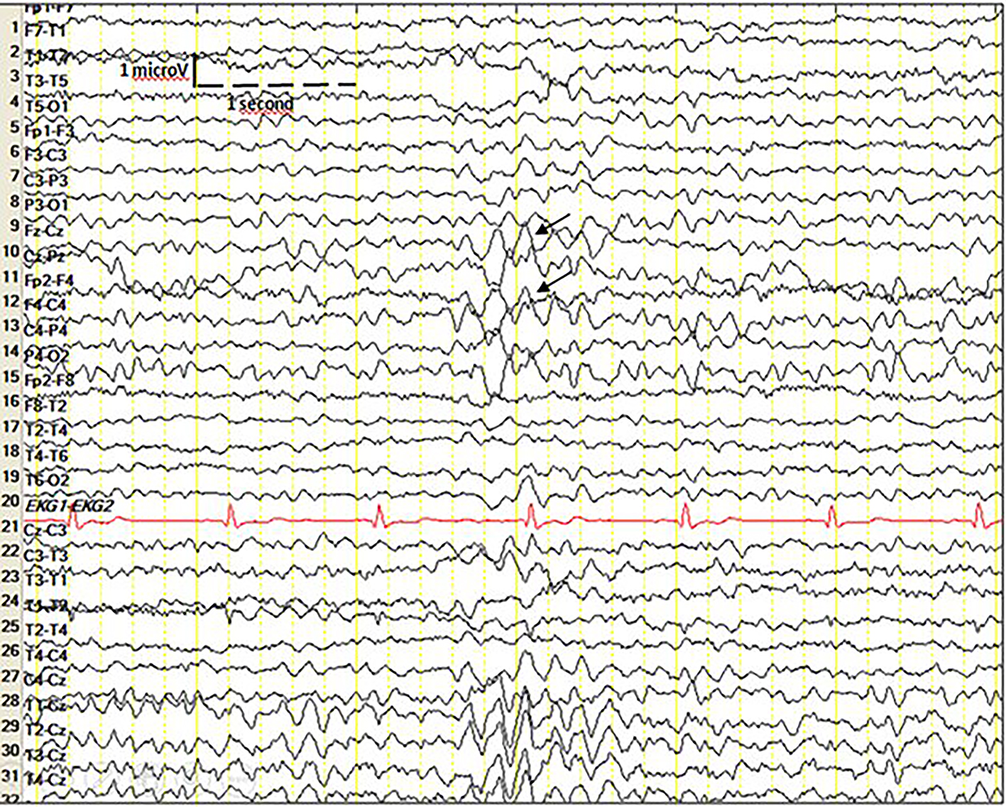
Figure 1. Routine EEG shows right centro-parietal paroxysmal disturbances characterized by sharp and slow waves. EEG: electroencephalogram.
| Journal of Neurology Research, ISSN 1923-2845 print, 1923-2853 online, Open Access |
| Article copyright, the authors; Journal compilation copyright, J Neurol Res and Elmer Press Inc |
| Journal website http://www.neurores.org |
Letter to the Editor
Volume 9, Number 3, June 2019, pages 48-50
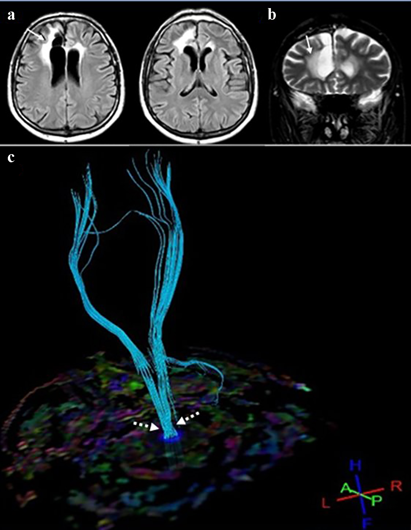
Detailed Illustration of a Patient With Ipsilateral Seizures
Figures