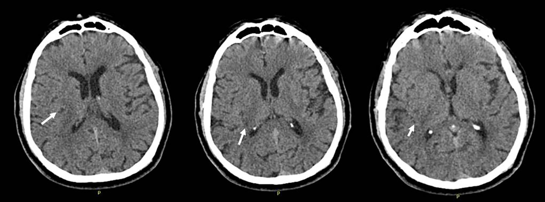
Figure 1. Cranial computerized tomography (CT) performed at initial admission, showing mild hypodensity in the right insula, parietal subcortical region.
| Journal of Neurology Research, ISSN 1923-2845 print, 1923-2853 online, Open Access |
| Article copyright, the authors; Journal compilation copyright, J Neurol Res and Elmer Press Inc |
| Journal website http://www.neurores.org |
Letter to the Editor
Volume 9, Number 1-2, March 2019, pages 18-20
Occam’s Razor Is Not Always True: Intracranial Tumor Mimicking Silent Infarction in a Patient With Previous Cerebral Infarction
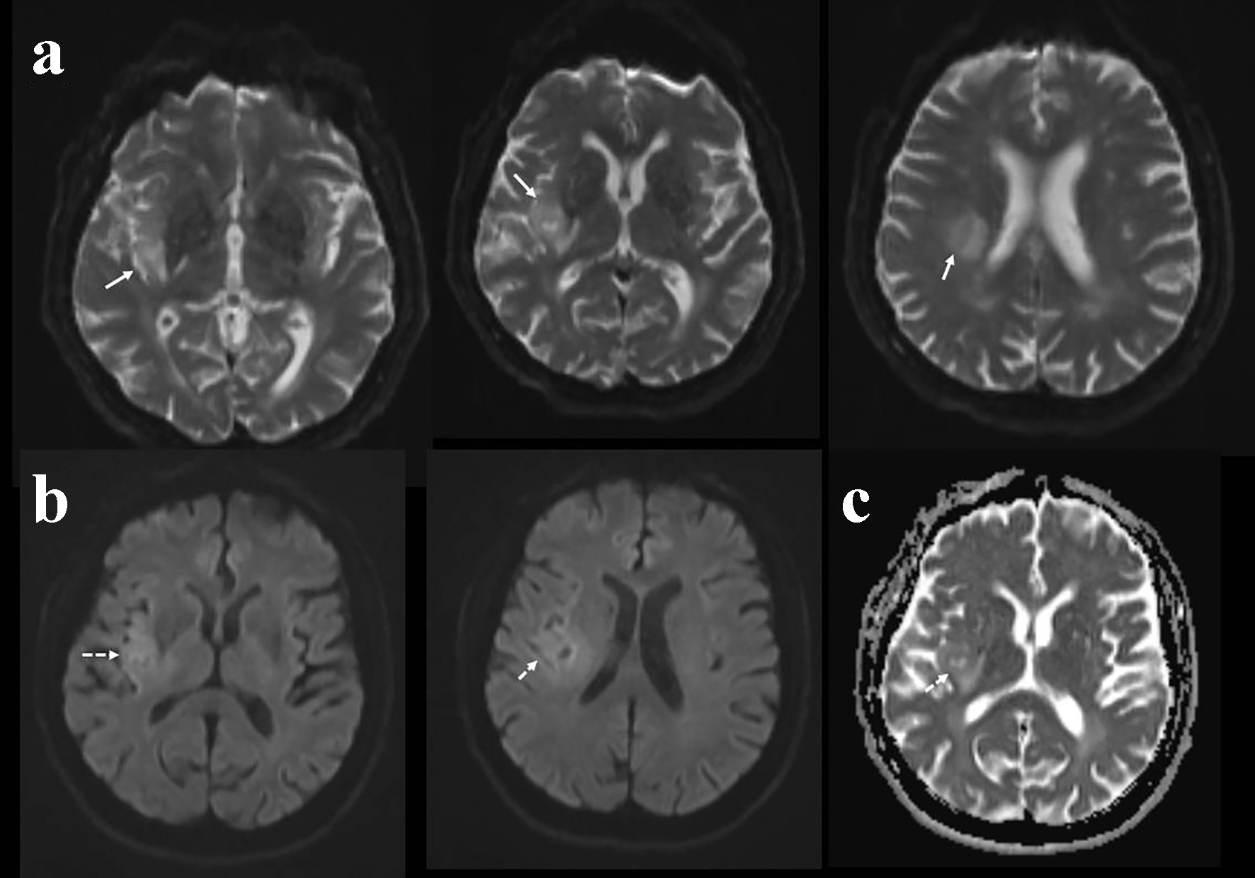
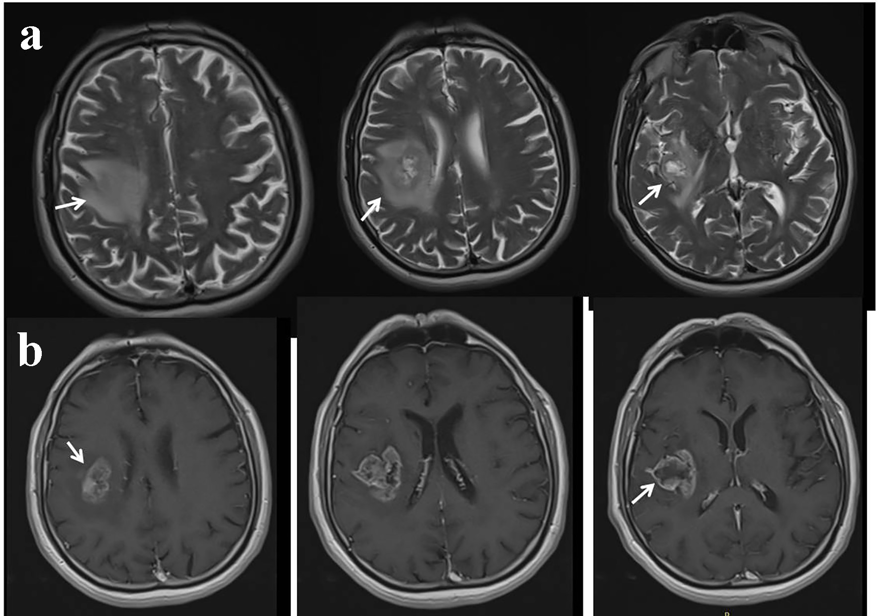
Figures