Journal of Neurology Research, ISSN 1923-2845 print, 1923-2853 online, Open Access
Article copyright, the authors; Journal compilation copyright, J Neurol Res and Elmer Press Inc
Journal website http://www.neurores.org

Case Report

Volume 7, Number 4-5, October 2017, pages 85-88


Chordoid Glioma of Third Ventricle Presenting as Primary Polydipsia With Aggressive Behavior: A Rare Case With Rare Presentation

Figures

Figure 1.
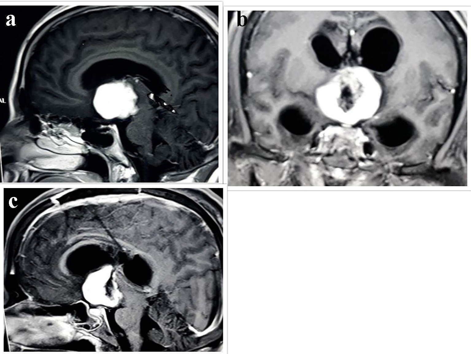
Figure 1. Radiological evaluation. (a) T1W MRI scan showing isointense lesion with homogenous enhancement in suprasellar and right parasellar region. Postoperative MRI scan after 2.5 months in (b) coronal and (c) sagittal scan showing heterogeneously enhancing hypointense mass lesion in suprasellar cistern and anterior part of third ventricle.
Figure 2.
Figure 2. Photomicrograph of chordoid glioma. (a) H&E stain shows cord like arrangement of epithelioid cells in myxomatous background. (b) Immunohistochemistry shows diffuse GFAP positivity. (c) EMA marks plasma cells. (d) CD34 shows patchy positivity.