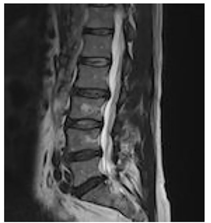
Figure 1. T2 axial MRI images show disc protrusion in L5-S1 level.
| Journal of Neurology Research, ISSN 1923-2845 print, 1923-2853 online, Open Access |
| Article copyright, the authors; Journal compilation copyright, J Neurol Res and Elmer Press Inc |
| Journal website http://www.neurores.org |
Case Report
Volume 7, Number 3, June 2017, pages 63-64
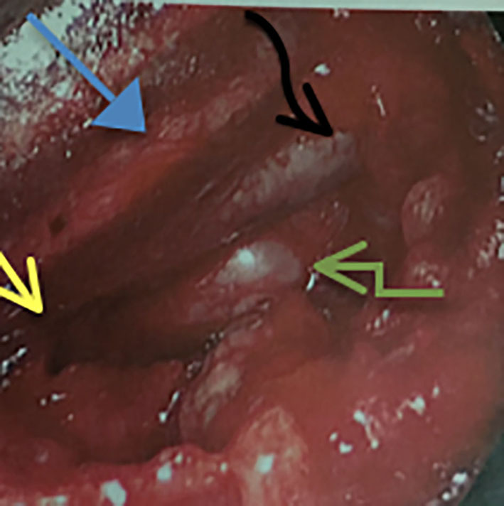
A Transverse Tri-Anastomotic Conjoined Nerve Root Discovered During a Lumbar Discectomy L5-S1
Figures