Journal of Neurology Research, ISSN 1923-2845 print, 1923-2853 online, Open Access
Article copyright, the authors; Journal compilation copyright, J Neurol Res and Elmer Press Inc
Journal website http://www.neurores.org

Case Report

Volume 3, Number 5, October 2013, pages 155-158


Optic Nerve Radiation Recall Associated With Prior Pemetrexed

Figures

Figure 1.
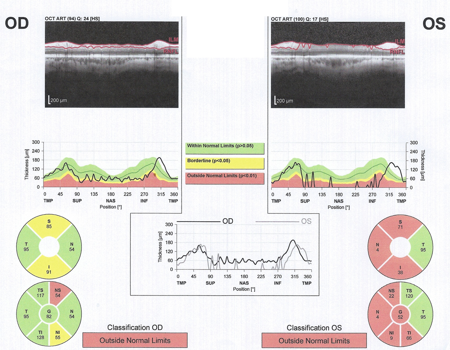
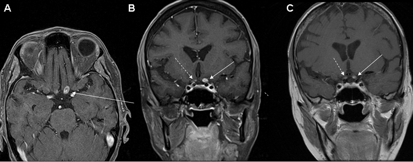
Figure 1. (A) Axial, and (B, C) coronal T1 post-contrast MRI images revealing an enhancing lesion of the posterior portion of the left optic nerve (solid arrow). There is also a smaller area of possible enhancement in the right optic nerve (dashed arrow) which is more clearly defined on the MRI (C) performed 6 weeks after the earlier study (B).
Figure 2.
Figure 2. Optical coherence tomography. Left (OS) greater than right (OD) retinal nerve fiber layer (RNFL) thinning is demonstrated.” In the current iteration the Left and Right are switched.