Journal of Neurology Research, ISSN 1923-2845 print, 1923-2853 online, Open Access
Article copyright, the authors; Journal compilation copyright, J Neurol Res and Elmer Press Inc
Journal website http://www.neurores.org

Case Report

Volume 3, Number 5, October 2013, pages 146-149


Abnormal Magnetic Resonance Imaging and Hemichorea Associated With Non-Ketotic Hyperglycemia

Figures

Figure 1.
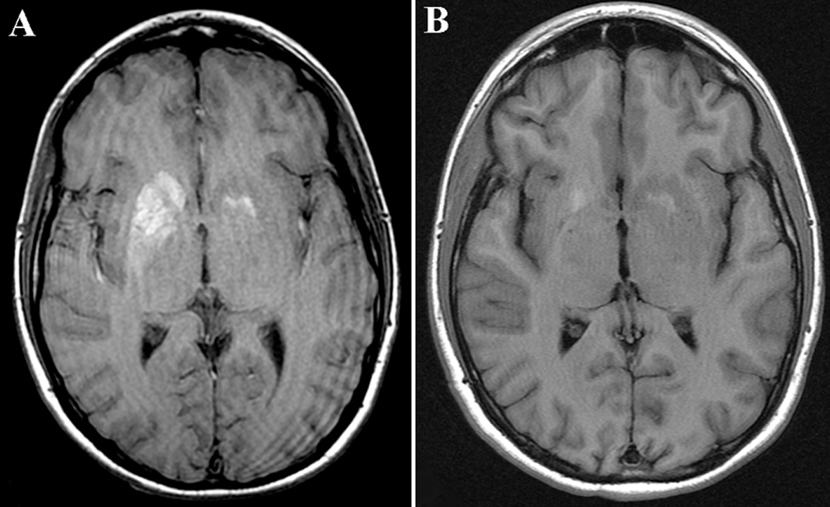
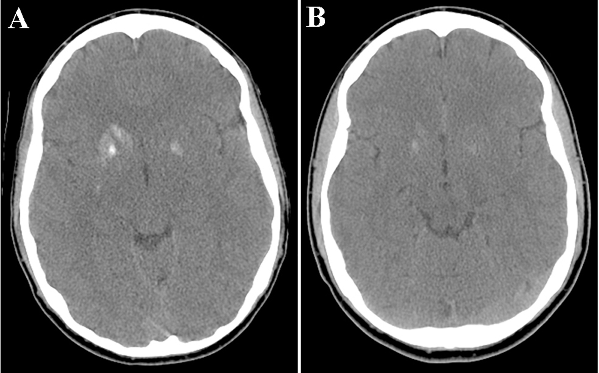
Figure 1. (A) A CT of the head without contrast demonstrated an area of hyperdensity involving the right head of caudate, putamen, and globus pallidus, and to a lesser extent, the left putamen, with no surrounding edema. (B) Partial resolution of the increased density in the right basal ganglia.
Figure 2.
Figure 2. (A) MRI T1-sequence of the brain showed an increased signal in the head of caudate and putamen on the right, and to a lesser extent, on the left. (B) Partial resolution of the increased signal abnormality in bilateral basal ganglia.
Figure 3.
Figure 3. An MRI of the brain with gadolinium revealed cortical enhancement in the right insula.