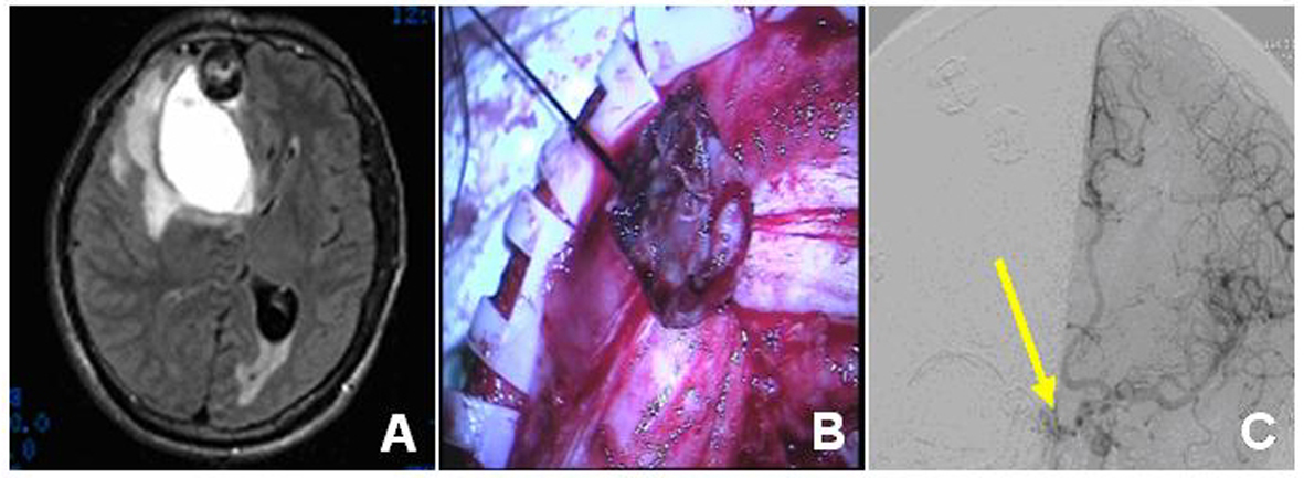
Figure 1. Imaging angiogram and intraoperative findings of case 1 patient. The preoperative CT (A) showed the right frontal lobe blood vessels and the nearby old hemorrhage. The intraoperative findings (B) revealed the aneurysmal dilatation with the purple membrane and the postoperative DSA showed the arteriovenous fistula artery stump (C, arrow).
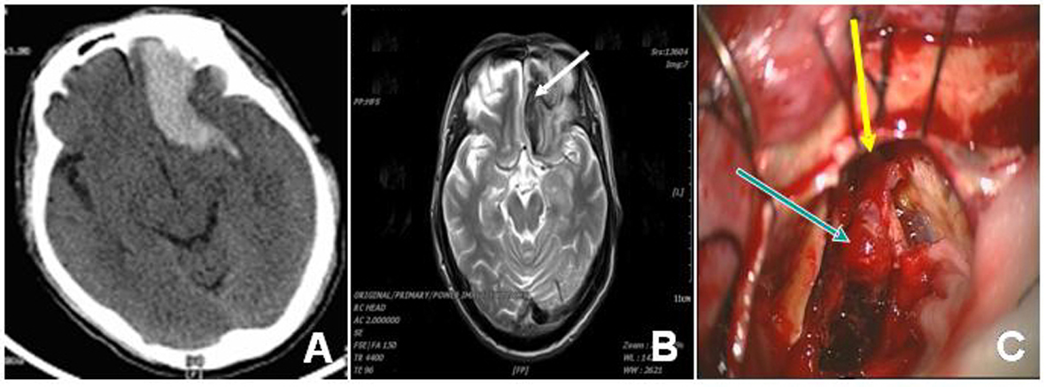
Figure 2. Imaging angiogram and intraoperative findings of case 2 patient. The preoperative CT (A) showed left frontal hemorrhage and the MRI T2-weighted image revealed hypointensity in the drainage vein (B, arrow). The intraoperative findings revealed the aneurysmal dilatation (C, arrow in blue) and its feeding artery (C, arrow in yellow).
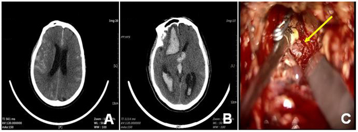
Figure 3. Imaging angiogram and intraoperative findings of case 3 patient. The original CT (A) showed SAH at cerebral base cistern and the preoperative CT (B) after re-admission revealed the hemorrhage in the brain ventricle. The intraoperative findings showed the aneurysmal dilatation (C, arrow).