Journal of Neurology Research, ISSN 1923-2845 print, 1923-2853 online, Open Access
Article copyright, the authors; Journal compilation copyright, J Neurol Res and Elmer Press Inc
Journal website http://www.neurores.org

Case Report

Volume 2, Number 4, August 2012, pages 176-179


A 12-Year-Old Girl With an Extraosseous Chondrosarcoma

Figures

Figure 1.
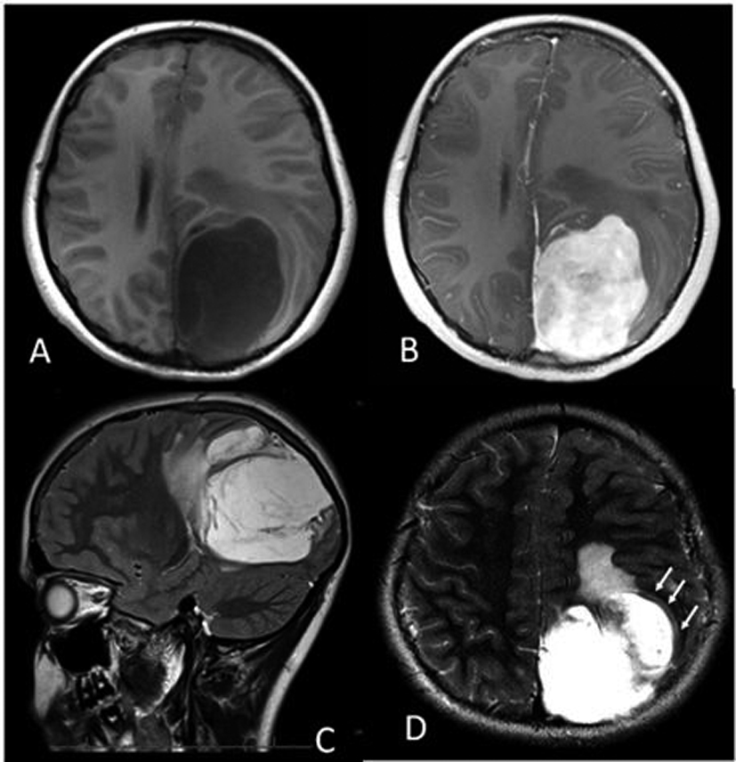
Figure 1. (A) Axial T1-weighted MRI showing the hypointense left parietal mass measuring approximately 6.5 × 5.0 × 7.0 cm, with 1.0 cm midline shift. (B) Axial post contrast T1-weighted MRI showing the heterogeneous, enhancing left parietal mass. (C) Sagittal T2-weighted image showing hyperintensity of the mass, with edema anteriorly. (D) Axial T2-weighted image depicting what is likely a CSF “cleft” (arrows), suggesting that the mass is extra-axial.
Figure 2.
Figure 2. (A) DWI MRI showing the left parietal mass without reduced diffusion. (B) ADC MRI showing the mass with abundant surrounding vasogenic edema, and internally high diffusivity (mean ADC = 2.221 × 10⁻³cm2/sec). For the purposes of comparison, a meningiosarcoma in a 17 year old is shown on post-contrast T1-weighted MRI (C), with ADC map (D) that has internally low diffusivity (asterisks, ADC = 0.677 × 10⁻³ cm2/sec).
Figure 3.
Figure 3. Hematoxylin and eosin staining showing the tumor with myxoid matrix and relatively low cellularity (Arrow showing myxoid matrix).