Journal of Neurology Research, ISSN 1923-2845 print, 1923-2853 online, Open Access
Article copyright, the authors; Journal compilation copyright, J Neurol Res and Elmer Press Inc
Journal website http://www.neurores.org

Case Report

Volume 2, Number 4, August 2012, pages 168-171


A Case of Amyloid-β-Related Angiitis: Histology, CSF-Findings and Treatment

Figures

Figure 1.
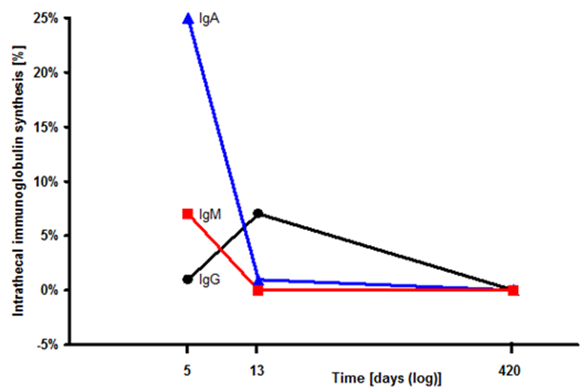
Figure 1. Intrathecal immunoglobulin synthesis in the cerebrospinal fluid (CSF): The CSF index for IgA (filled triangles), IgM (filled squares), IgG (filled circles) successively declined during steroid therapy.
Figure 2.
Figure 2. Axial Cranial MRI: Susceptibility Weighted Imaging (SWI) MRI displays multifocal black dots localized in the left parietooccipital lobe (A). Fluid Attenuated Inversion Recovery (FLAIR) MRI shows multiple white matter lesions particularly in the left parietooccipital area with an extensive edema and significant mass effect (B).
Figure 3.
Figure 3. Brain biopsy (left occipital lobe): A medium sized vessel with granulomatous inflammatory infiltrate, deposition of amyloid-β in vessel wall (A, immunohistochemical staining against amyloid-β, arrow) and multinucleated macrophages (B, immunohistochemical staining with the macrophage marker CD68, arrow).