Journal of Neurology Research, ISSN 1923-2845 print, 1923-2853 online, Open Access
Article copyright, the authors; Journal compilation copyright, J Neurol Res and Elmer Press Inc
Journal website http://www.neurores.org

Case Report

Volume 2, Number 4, August 2012, pages 164-167


Spinal Hemorrhage Leading to Cerebral Vasospasm and Infarction

Figures

Figure 1.
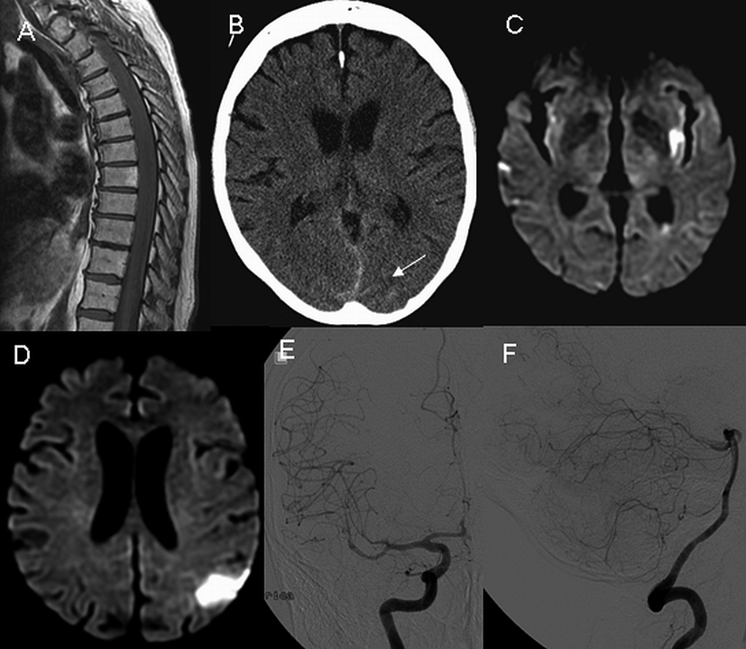
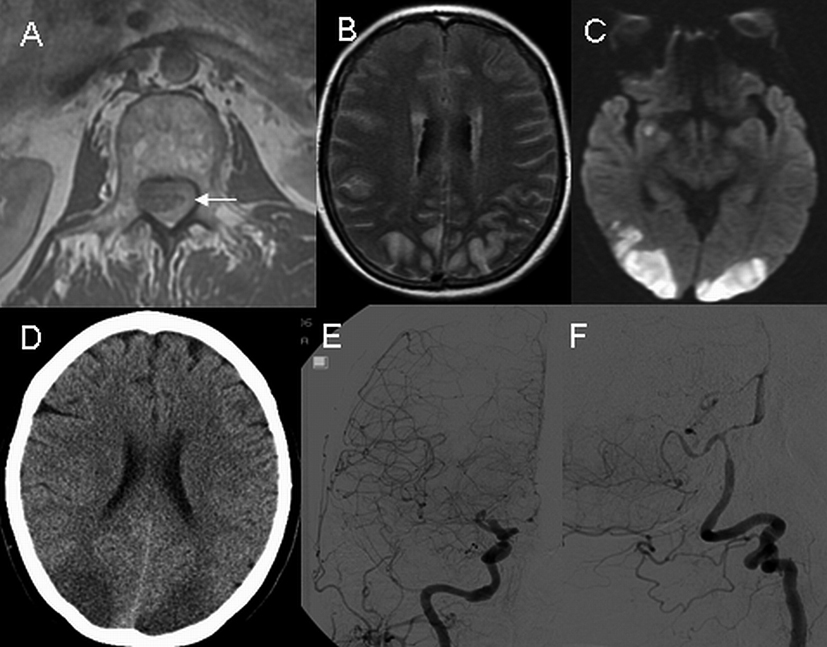
Figure 1. Axial T1- weighted MRI of the upper lumbar spine on day 11 shows a spinal hemorrhage (A, arrow). On day 14, axial MRI images of the head reveal diffusely increased T2 signals in the subarachnoid space (B) and presence of ischemia on diffusion-weighted imaging (C). On day 15, non-contrast CT scan of head reveals ischemia without hemorrhage (D), and digital subtraction angiography demonstrates diffuse narrowing of the carotid (E) and vertebrobasilar (F) arteries.
Figure 2.
Figure 2. Sagittal T1 weighted MRI of the thoracic spine reveals extensive hemorrhage on day 5 (A). Non-contrast head CT on day 16 shows the presence of minimal amount of subarachnoid hemorrhage (B, arrow). On day 20, axial diffusion-weighted images of the head demonstrate multiple areas of ischemia (C and D). Digital subtraction angiography on day 22 demonstrates the presence of diffuse narrowing of the carotid (E) and vertebrobasilar (F) arteries.