Journal of Neurology Research, ISSN 1923-2845 print, 1923-2853 online, Open Access
Article copyright, the authors; Journal compilation copyright, J Neurol Res and Elmer Press Inc
Journal website http://www.neurores.org

Case Report

Volume 2, Number 3, June 2012, pages 123-126


Embolic Brain Abscess in a Patient With Tricuspid Valve Endocarditis and a Patent Foramen Ovale

Figures

Figure 1.
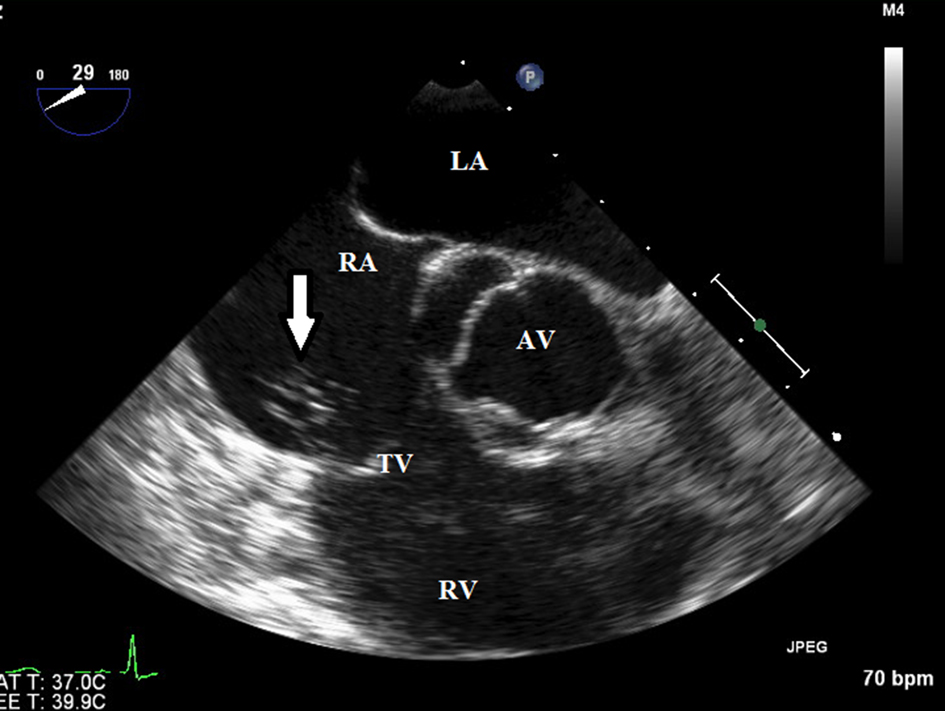
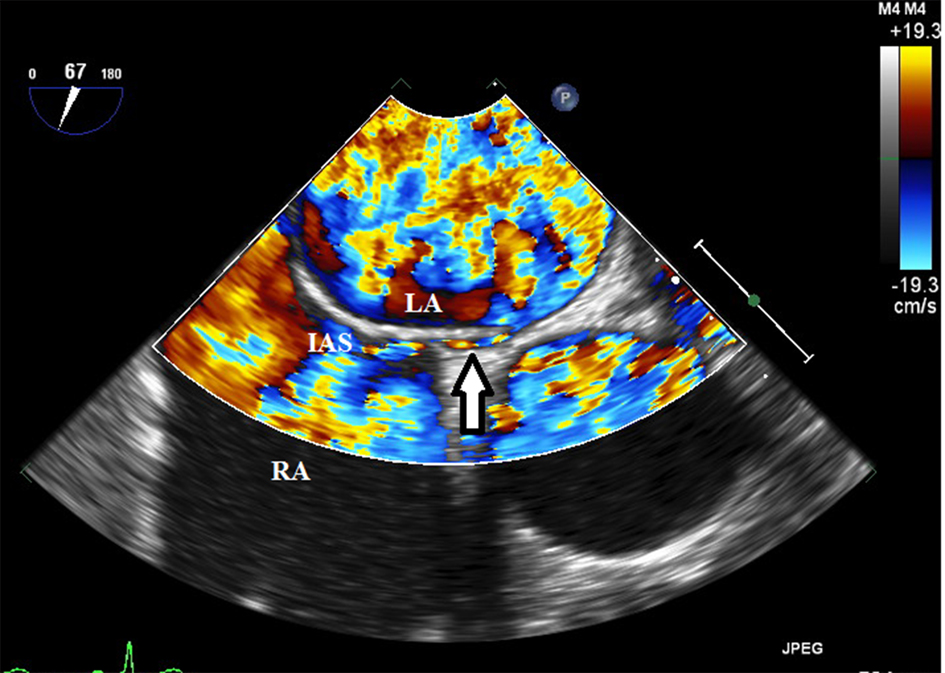
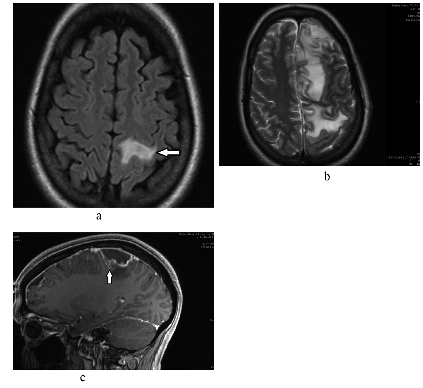
Figure 1. a. Brain MRI shows 1.1 x 1.1 x 1.0 cm peripherally enhancing lesion (arrow) in the superior aspect of the left precentral gyrus with surrounding edema and mild enhancement of the surrounding dura. b. Repeat brain MRI shows a worsening appearance with thickened enhancing pachymeninges of the superior frontal and parietal region as well as diffuses leptomeningeal enhancement and increasing mass effect. There was significant increased abnormal T2 signal throughout the superior left cerebral hemisphere consistent with vasogenic edema. c. Midline sagittal cut showing brain abscess (arrow) with significant surrounding edema.
Figure 2.
Figure 2. Transesopagheal echocardiogram showed a large amorphous mobile echodensity arising from the anterior tricuspid valve leaflet (arrow) highly suspicious for a vegeta.
Figure 3.
Figure 3. A small Patent foramen ovale (arrow) was demonstrated by color Doppler and agitated saline contrast injection. LA: left atrium; RA: right atrium; IAS: interatrial septum; AV: aortic valve; TV: tricuspid valve.