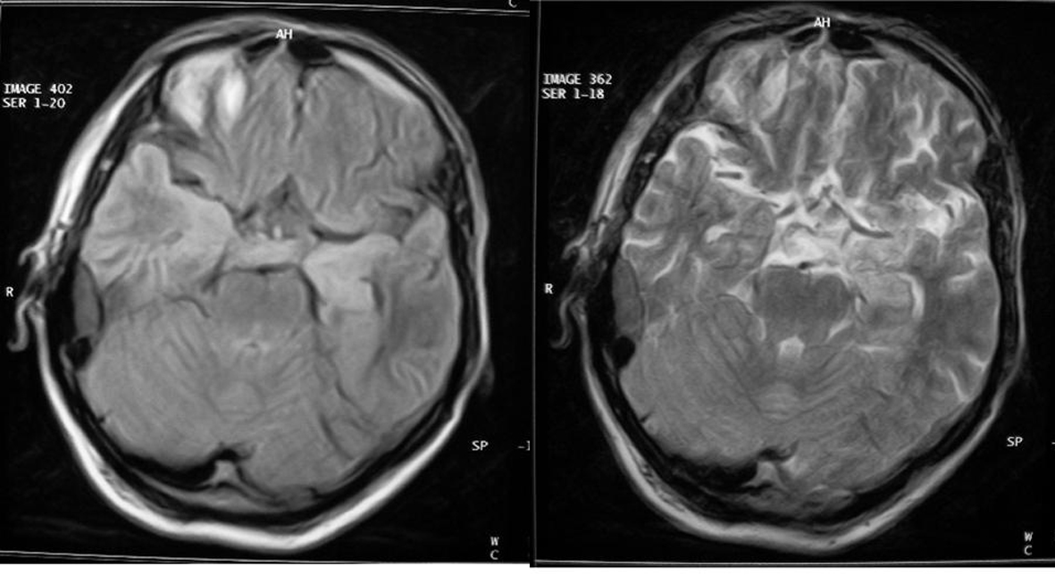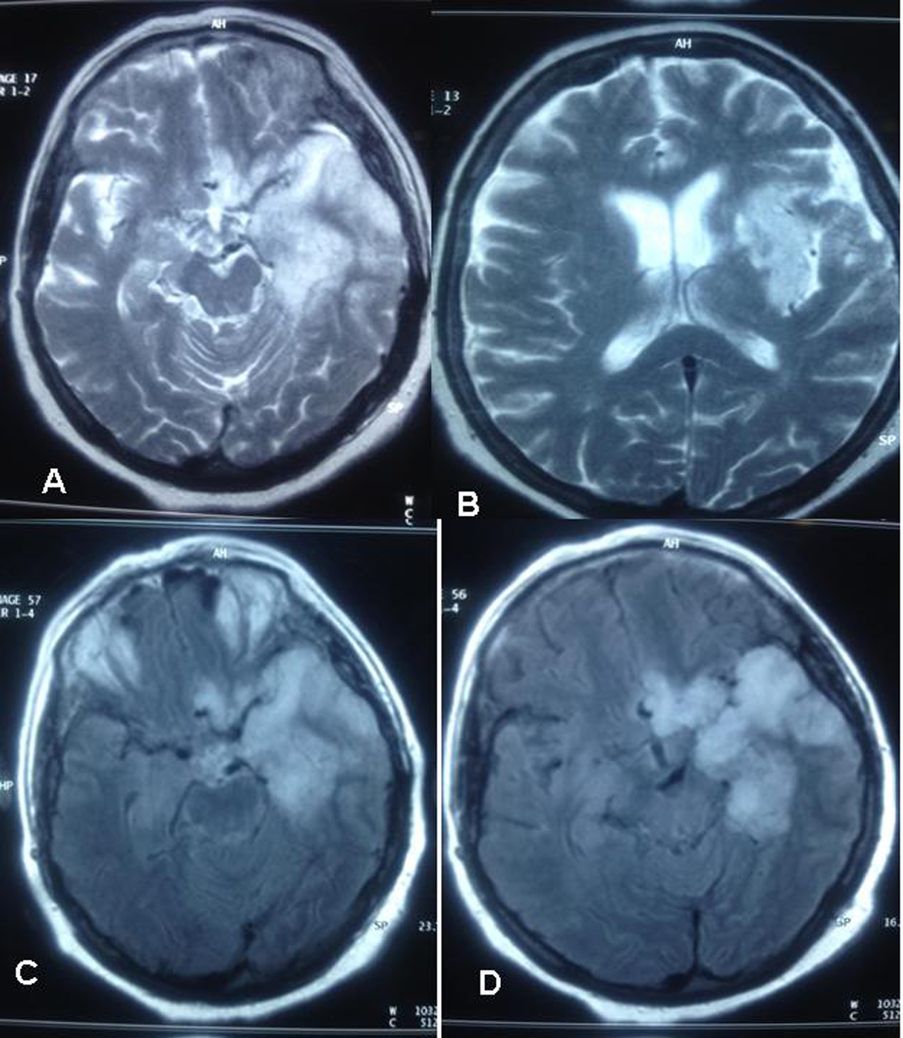
Figure 1. Cranial MRI, lesions were seen more clearly in the left temporal region and as increased intensity in hypothalamus and insula region.
| Journal of Neurology Research, ISSN 1923-2845 print, 1923-2853 online, Open Access |
| Article copyright, the authors; Journal compilation copyright, J Neurol Res and Elmer Press Inc |
| Journal website http://www.neurores.org |
Case Report
Volume 2, Number 3, June 2012, pages 104-108
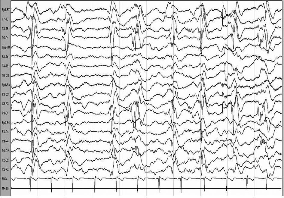
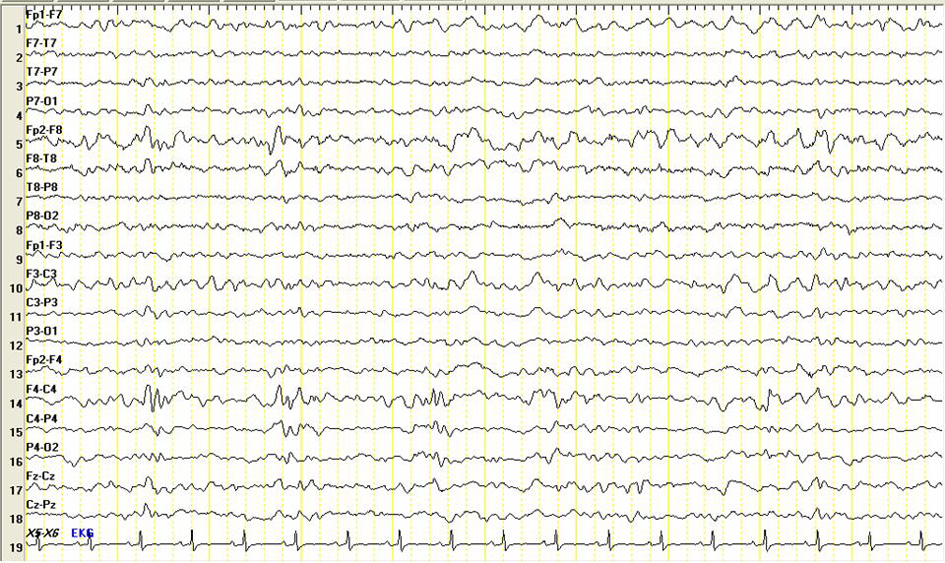
Presenting With Status Epilepticus That Neurosyphilis: Case Report
Figures