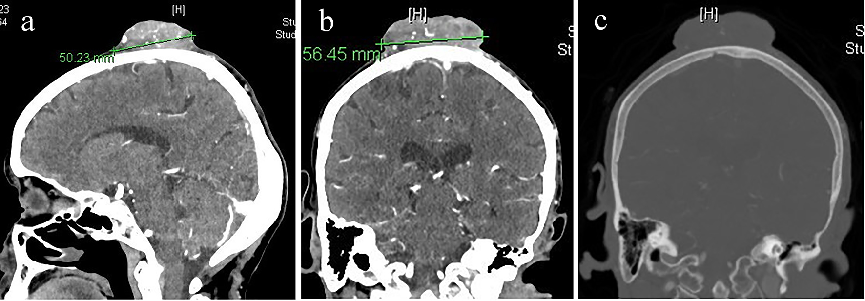
Figure 1. (a) CT of brain sagittal cut with IV contrast. (b) CT of brain coronal cut with IV contrast showing moderately vascular scalp mass. (c) CT of brain bone window showing no bone invasion or intracranial extension.
| Journal of Neurology Research, ISSN 1923-2845 print, 1923-2853 online, Open Access |
| Article copyright, the authors; Journal compilation copyright, J Neurol Res and Elmer Press Inc |
| Journal website http://www.neurores.org |
Case Report
Volume 7, Number 6, December 2017, pages 115-117
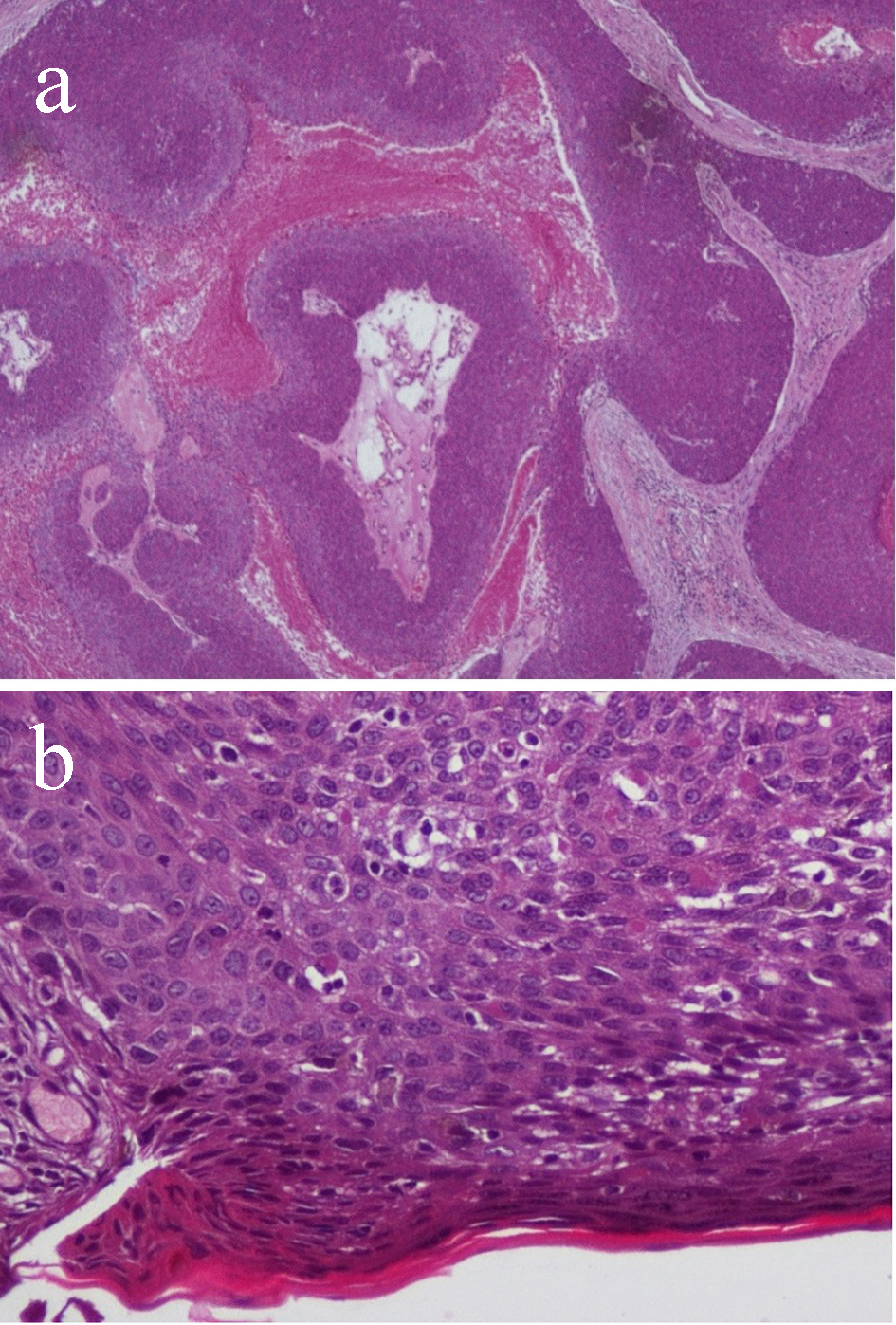
Eccrine Porocarcinoma of the Scalp: A Case Report and Review of Literature
Figures