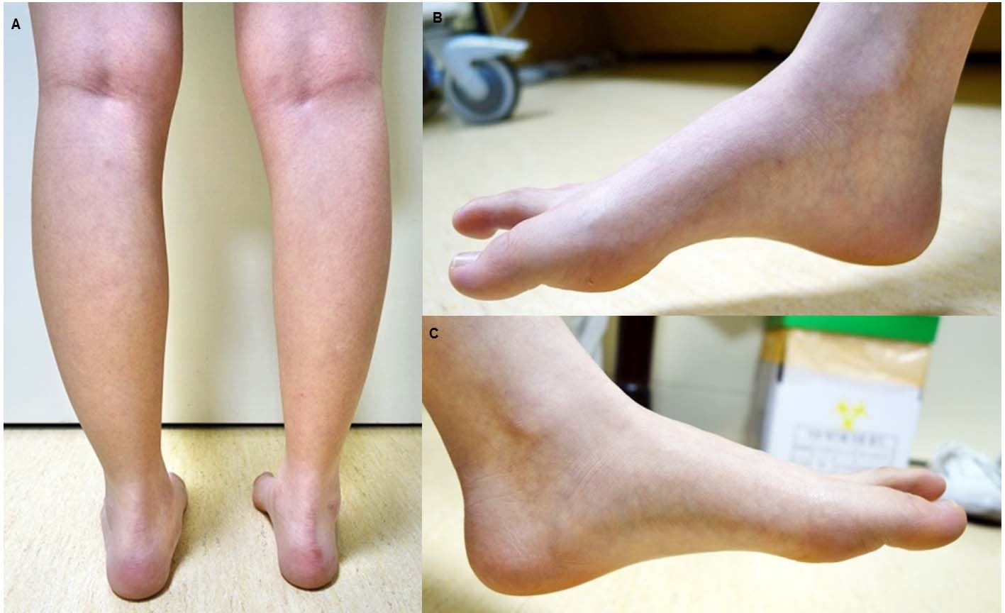
Figure 1. Photographs of lower limbs of patient show marked muscle atrophy of posterior compartment of right calf (A) and evidence of pes cavus (B). The left plantar muscle is normal (C).
| Journal of Neurology Research, ISSN 1923-2845 print, 1923-2853 online, Open Access |
| Article copyright, the authors; Journal compilation copyright, J Neurol Res and Elmer Press Inc |
| Journal website http://www.neurores.org |
Case Report
Volume 6, Number 5-6, December 2016, pages 118-123
Suspected Anterior Horn Disease Mistaken as a Mass in the Sacral Plexus: A Case Report
Figures
Tables
| Latency (ms) | Conduction velocity (m/s) | Amplitude (motor, mV/sensory, μV) | |||||||
|---|---|---|---|---|---|---|---|---|---|
| RT | LT | NL | RT | LT | NL | RT | LT | NL | |
| RT: right; LT: left; NL: normal range; NR: no response; EDB: extensor digitorum brevis; B. Fib: below fibular head; Poplit: popliteal fossa; Fib head: fibular head. | |||||||||
| Initial | |||||||||
| Motor nerve | |||||||||
| Peroneal nerve | |||||||||
| EDB | |||||||||
| Ankle | 3.1 | 2.7 | ≤ 6.5 | 7.4 | 8.8 | ≥ 4 | |||
| B. Fib | 9.0 | 8.3 | 47 | 50 | ≥ 41.8 | 7.0 | 8.0 | ||
| Poplit | 10.5 | 9.4 | 47 | 51 | ≥ 40.0 | 6.0 | 7.8 | ||
| Tibial nerve | |||||||||
| Ankle | 8.8 | 3.2 | ≤ 5.8 | 2.8 | 24.6 | ≥ 5 | |||
| Knee | 18.9 | 9.7 | 35 | 55 | ≥ 40.6 | 1.9 | 23.3 | ||
| Plantar | |||||||||
| Medial | 8.4 | 3.0 | ≤ 5.4 | 3.3 | 36.5 | ≥ 3.5 | |||
| Lateral | 8.2 | 4.3 | ≤ 6.3 | 1.8 | 19.4 | ≥ 3.0 | |||
| Sensory nerve | |||||||||
| Lateral femoral cutaneous nerve | 2.8 | 2.5 | ≤ 3.0 | 61 | 58 | ≥ 40.3 | 11.8 | 13.6 | ≥ 2 |
| Peroneal nerve | 2.8 | 2.8 | ≤ 4.4 | 48.0 | 50 | ≥ 40.5 | 48 | 38.1 | ≥ 4.0 |
| Sural nerve | 3.4 | 3.3 | ≤ 4.4 | 35 | 35 | ≥ 34.6 | 14.4 | 14.8 | ≥ 6.0 |
| Plantar | |||||||||
| Medial | 2.3 | 2.9 | 10.6 | 12.1 | |||||
| Lateral | 2.4 | 3.0 | 11.2 | 13.7 | |||||
| After 11 months | |||||||||
| Motor nerve | |||||||||
| Peroneal nerve | |||||||||
| EDB | |||||||||
| Ankle | 4.8 | 2.8 | ≤ 6.5 | 9.2 | 10.7 | ≥ 4 | |||
| B. Fib | 11.1 | 8.5 | 48 | 53 | ≥ 41.8 | 8.6 | 10.5 | ||
| Poplit | 12.4 | 9.4 | 54 | 67 | ≥ 40.0 | 8.5 | 10.5 | ||
| Tibial nerve | |||||||||
| Ankle | 8.8 | 3.5 | ≤ 5.8 | 2.7 | 24.6 | ≥ 5 | |||
| Knee | 18.9 | 9.9 | 35 | 55 | ≥ 40.6 | 1.9 | 23.3 | ||
| Plantar | |||||||||
| Medial | 8.4 | 3.0 | ≤ 5.4 | 3.2 | 36.5 | ≥ 3.5 | |||
| Lateral | 8.2 | 4.3 | ≤ 6.3 | 1.8 | 19.4 | ≥ 3.0 | |||
| Sensory nerve | |||||||||
| Lateral femoral cutaneous nerve | 3.1 | 3.0 | ≤ 3.0 | 61 | 58 | ≥ 40.3 | 11.8 | 13.6 | ≥ 2 |
| Peroneal nerve | 2.7 | 2.9 | ≤ 4.4 | 52 | 48 | ≥ 40.5 | 9.1 | 10.8 | ≥ 4.0 |
| Sural nerve | 3.6 | 3.2 | ≤ 4.4 | 35 | 38 | ≥ 34.6 | 19.3 | 12.1 | ≥ 6.0 |
| Plantar | |||||||||
| Medial | 2.4 | 2.9 | 10.2 | 12.1 | |||||
| Lateral | 2.3 | 3.0 | 10.8 | 13.7 | |||||
| Muscle | Insertional activity | Fibrillation | Positive sharp wave | MUAP | IP |
|---|---|---|---|---|---|
| RT: right; LT: left; MUAP: motor unit action potential; IP: interference pattern; NP: non-production of MUAP; RIP: reduced interference pattern; FIP: full interference pattern. | |||||
| Gluteus maximus | |||||
| RT | Normal | - | - | Normal | FIP |
| LT | Normal | - | - | Normal | FIP |
| Gluteus medius | |||||
| RT | Normal | - | - | Normal | FIP |
| LT | Normal | - | - | Normal | FIP |
| Vastus lateralis | |||||
| RT | Normal | - | - | Normal | FIP |
| LT | Normal | - | - | Normal | FIP |
| Biceps femoris short and long head | |||||
| RT | Normal | - | - | Normal | FIP |
| LT | Normal | - | - | Normal | FIP |
| Semimembranosus | |||||
| RT | ↑ | + | ++ | Increased | RIP |
| LT | Normal | - | - | Normal | FIP |
| Semitendinosus | |||||
| RT | ↑ | - | ++ | Normal | RIP |
| LT | Normal | - | - | Normal | FIP |
| Tibialis anterior | |||||
| RT | Normal | - | - | Normal | FIP |
| LT | Normal | - | - | Normal | FIP |
| Peroneus longus | |||||
| RT | Normal | - | - | Normal | FIP |
| LT | Normal | - | - | Normal | FIP |
| Tibialis posterior | |||||
| RT | ↑ | + | ++ | Increased | RIP |
| LT | Normal | - | - | Normal | FIP |
| Gastrocnemius medius | |||||
| RT | ↑ | + | ++ | Increased | RIP |
| LT | Normal | - | - | Normal | FIP |
| Abductor halluces brevis | |||||
| RT | ↑ | - | ++ | NP | - |
| LT | Normal | - | - | Normal | FIP |
| Abductor digiti quinti pedis | |||||
| RT | ↑ | - | ++ | NP | - |
| LT | Normal | - | - | Normal | FIP |
| Paraspinal (C4-T1, L1-S2) | |||||
| RT | Normal | - | - | - | - |
| LT | Normal | - | - | - | - |