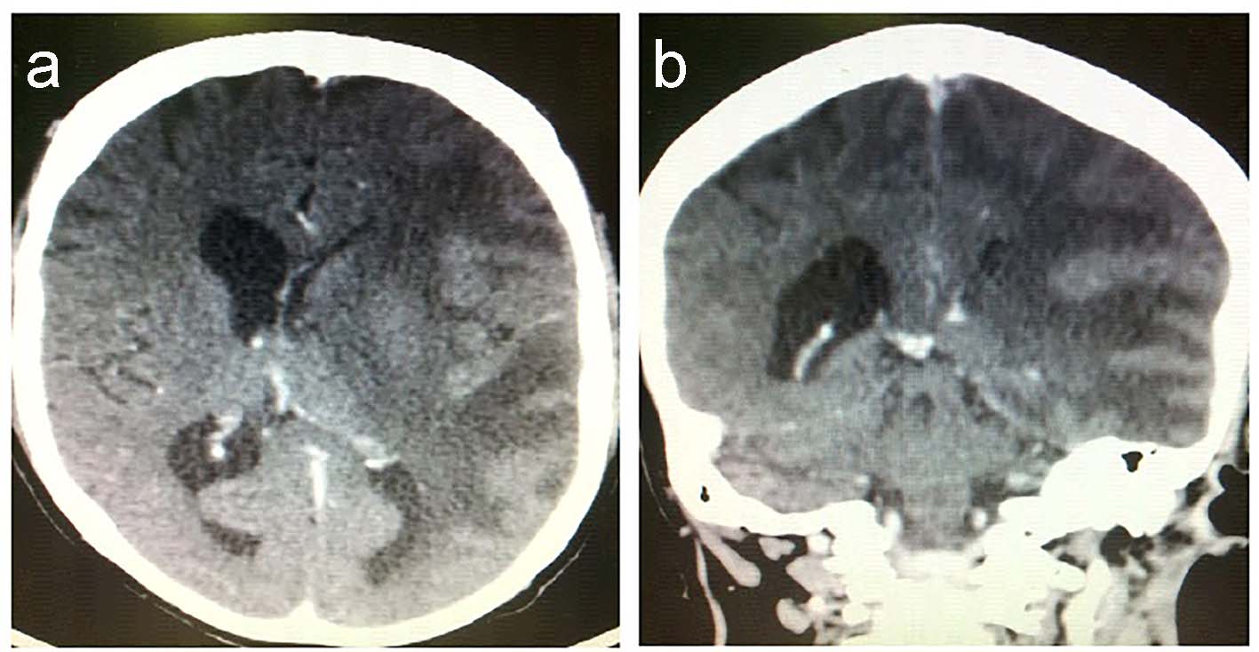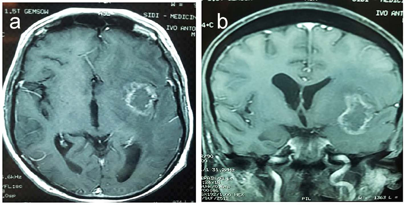
Figure 1. Enhanced T1WI MRI with a left insular lesion: (a) axial view; (b) coronal view.
| Journal of Neurology Research, ISSN 1923-2845 print, 1923-2853 online, Open Access |
| Article copyright, the authors; Journal compilation copyright, J Neurol Res and Elmer Press Inc |
| Journal website http://www.neurores.org |
Case Report
Volume 6, Number 5-6, December 2016, pages 102-105
Brain Radionecrosis: Case Report
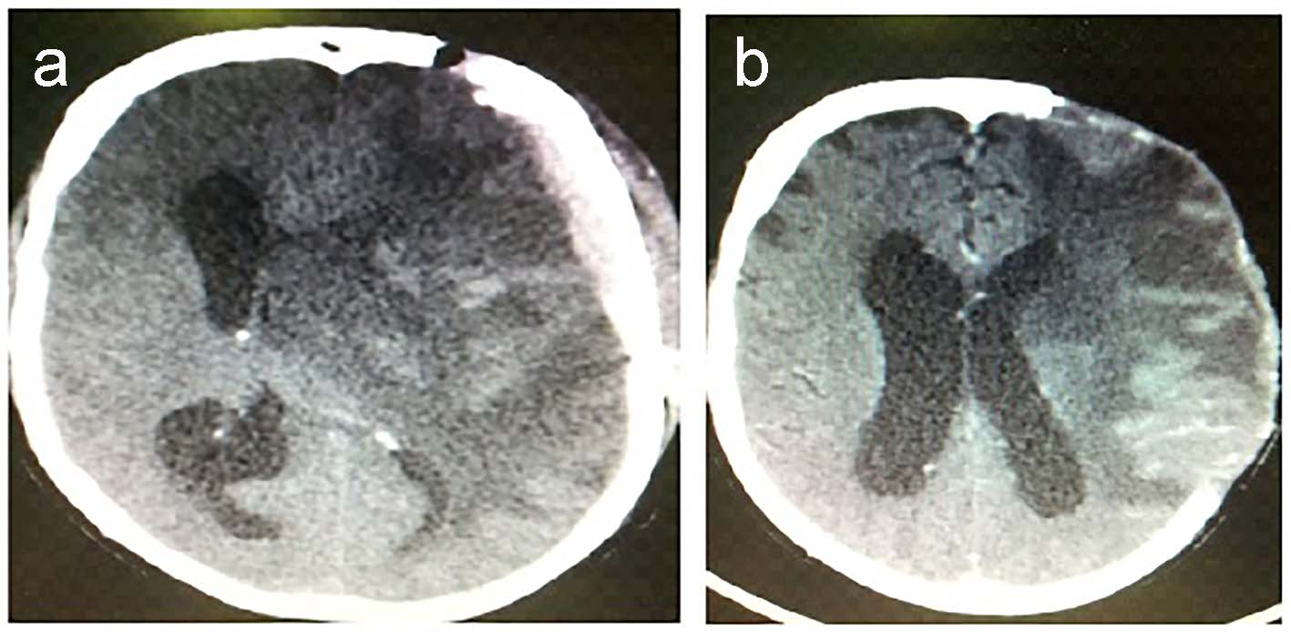
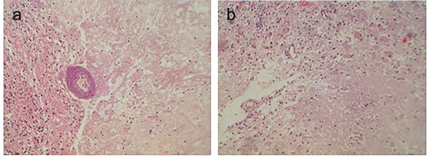
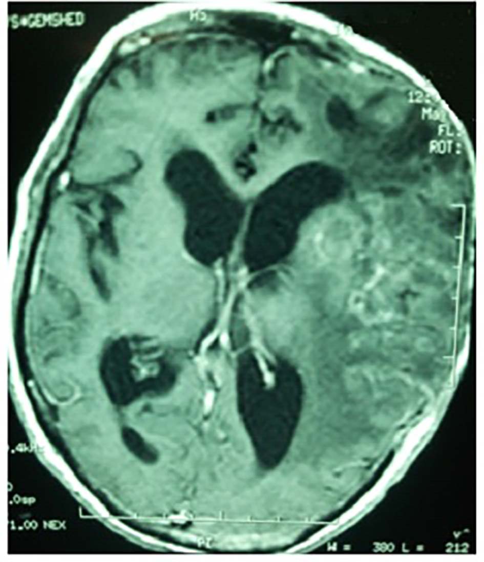
Figures