Journal of Neurology Research, ISSN 1923-2845 print, 1923-2853 online, Open Access
Article copyright, the authors; Journal compilation copyright, J Neurol Res and Elmer Press Inc
Journal website http://www.neurores.org

Case Report

Volume 6, Number 2-3, June 2016, pages 53-56


Acute Disseminated Encephalopathy Combined With Anticentromere Antibody B Positive: Uncommon Primary Manifestations of Sjogren’s Syndrome

Figures

Figure 1.
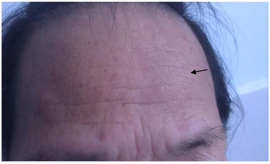
Figure 1. Forehead pachylosis (arrow).
Figure 2.
Figure 2. Brain CT obtained on the day of admission. The images show multiple hypodensity lesions in the bilateral hemispheres.
Figure 3.
Figure 3. The first brain MRI after admission. (a) The sequence of T2-weighted image showed multiple hyperdensity round lesions in the bilateral hemispheres, mainly involved the white matter, which were surrounded by schistose brain edema. (b) The sequence of T1-weighted image revealed that the lesions were hypodensity. (c) The enhanced sequence showed looping and nodal enhancement. (d) DWI prompted some lesions were hyperdensity, which revealed restriction of diffusion. (e) MRA showed feckly normal.
Figure 4.
Figure 4. Repeated brain MRI revealed that most of the lesions and hydrocephalus were remitted.
Figure 5.
Figure 5. Labial salivary gland biopsy showed two foci of focal lymphocytic sialadenitis.