Journal of Neurology Research, ISSN 1923-2845 print, 1923-2853 online, Open Access
Article copyright, the authors; Journal compilation copyright, J Neurol Res and Elmer Press Inc
Journal website http://www.neurores.org

Case Report

Volume 6, Number 1, February 2016, pages 24-27


Anterior Choroidal Artery Aneurysm Associated With Moyamoya Disease

Figures

Figure 1.
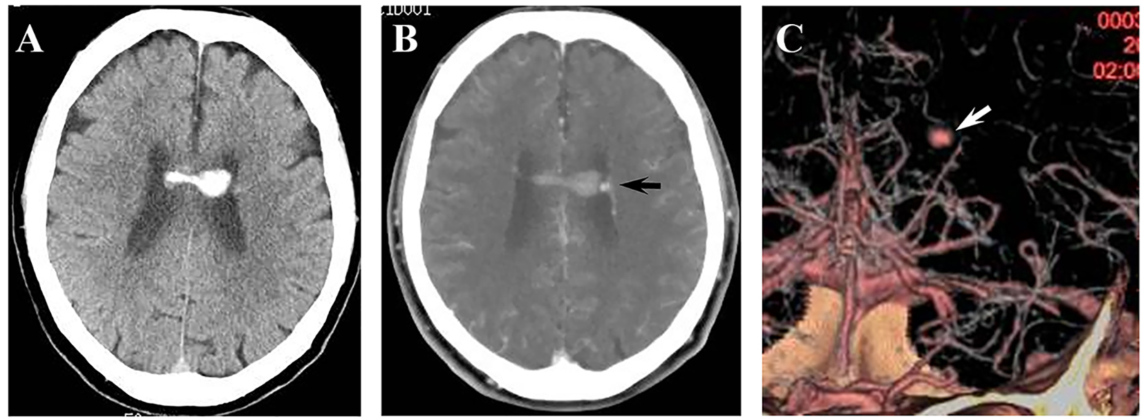
Figure 1. (A) CT on admission showing intraventricular hemorrhage. (B) CECT showing an enhanced aneurysm in the left lateral ventricle (arrow). (C) 3D-CTA showing an aneurysm on the left AChoA (arrow).
Figure 2.
Figure 2. (A) MRI obtained 13 months before admission showing no aneurysm. (B) MRI on admission showing an aneurysm on AChoA (arrow). (C) MRI on the 17th day showing disappearance of the AChoA aneurysm. (D) MRA obtained 8 months after surgery, showing no recurrence of the aneurysm.
Figure 3.
Figure 3. (A) Angiography on admission showing an aneurysm of 4 mm in diameter on the left AChoA (arrow). (B) Angiography on the ninth day showing enlargement of the aneurysm. (C) Angiography on the 17th day showing disappearance of the aneurysm. (D) Angiography obtained 13 months after onset showing patency of the bypass and no opacification of the aneurysm.