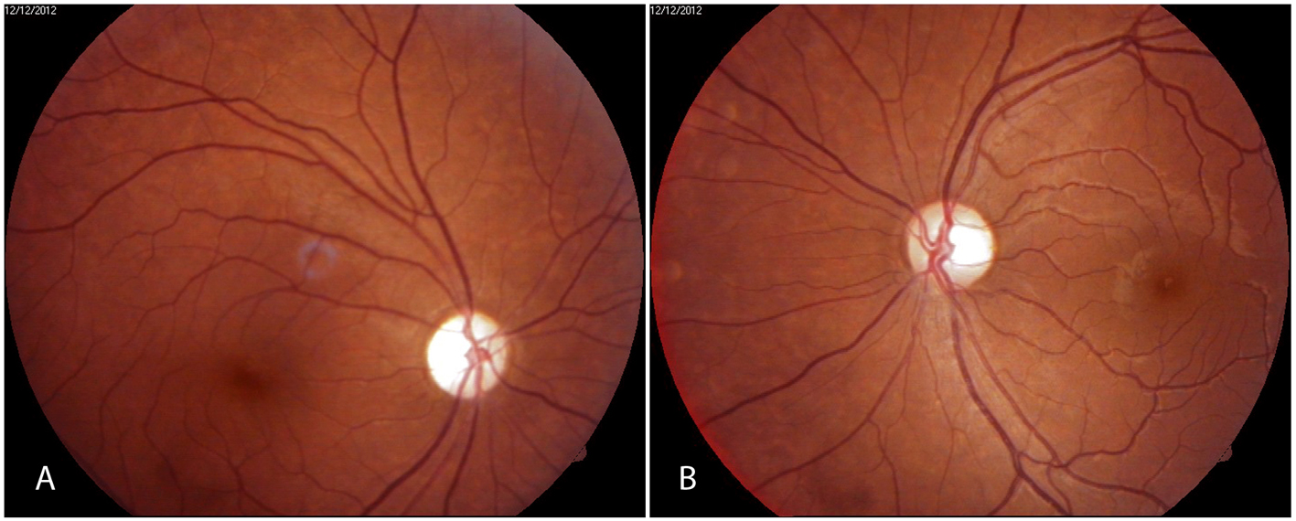
Figure 1. Fundus photographs showing (A) optic atrophy right eye and (B) temporal pallor of optic disk in left eye.
| Journal of Neurology Research, ISSN 1923-2845 print, 1923-2853 online, Open Access |
| Article copyright, the authors; Journal compilation copyright, J Neurol Res and Elmer Press Inc |
| Journal website http://www.neurores.org |
Case Report
Volume 4, Number 5-6, December 2014, pages 145-149
Ocular Oscillations and Transient Oscillopsia in Neuromyelitis Optica
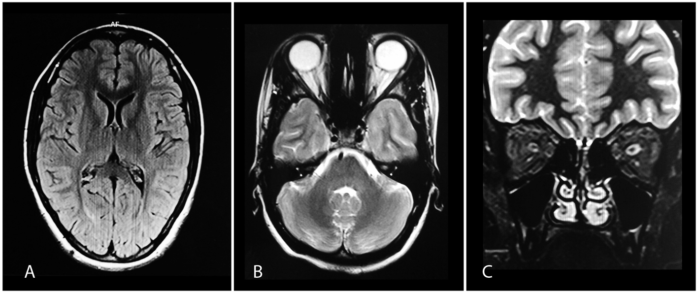
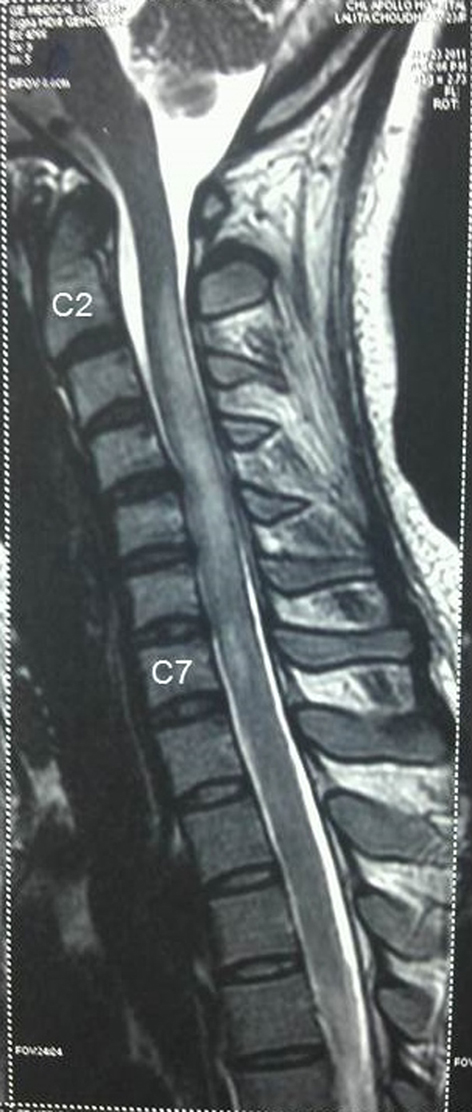
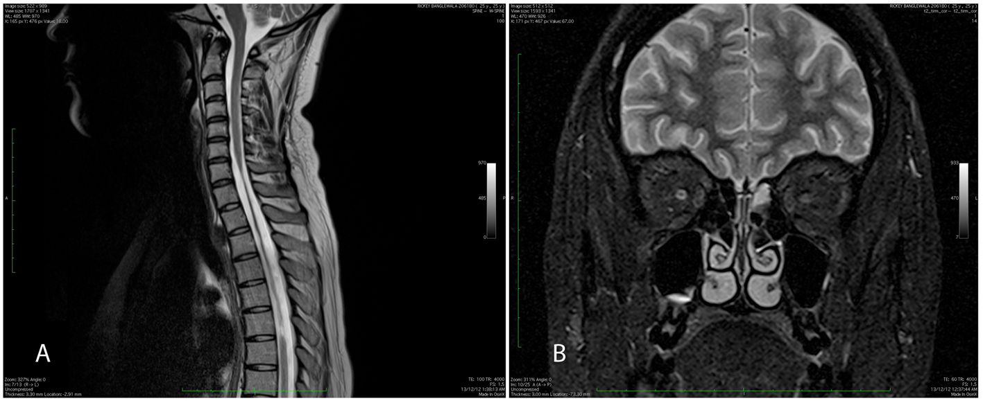
Figures