Journal of Neurology Research, ISSN 1923-2845 print, 1923-2853 online, Open Access
Article copyright, the authors; Journal compilation copyright, J Neurol Res and Elmer Press Inc
Journal website http://www.neurores.org

Original Article

Volume 4, Number 5-6, December 2014, pages 132-137


Key Role of Promoter Methylation and Inactivation of MCPH1 Gene in Brain Tumors

Figures

Figure 1.
Figure 1. (a) MSP-PCR analysis of brain tumor tissue samples. M: marker (50 bp ladder); u65: PCR using specific primers for unmethylated template 65 (201 bps); m2: PCR using specific primers for methylated template 2(203 bps). (b) Sequencing analysis of MSP-PCR of methylated template.
Figure 2.
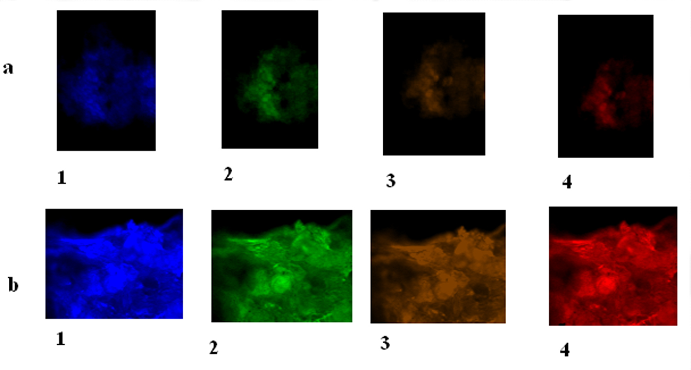
Figure 2. MCPH1 protein expression of cyclin E, CDC25A and MPCH1 in astrocytoma and meningioma tumors. (a) Brain tumor cells (BTCs) with astrocytoma. 1: BTC with DAPI filter; 2: BTC conjugated with FITC presenting low expression of cyclin E; 3: BTC conjugated with R-PE presenting low expression of CDC25A; 4: BTC conjugated with PE-Cy5 reflecting low expression of MCPH1 (× 100). (b) BTCs with meningioma. 1: BTC with DAPI filter; 2: BTC conjugated with FITC presenting high expression of cyclin E; 3: BTC conjugated with R-PE presenting low expression of cyclin E in majority of cells accompanied by clone of cells with high expression; 4: BTC conjugated with PE-Cy5 reflecting high expression of MCPH1 (× 100).
Figure 3.
Figure 3. Q-FISH illustrates status of telomere of an astrocytoma tumor in a patient affected with primary brain tumor. (a) DAPI filter; (b) Cy3 showing tumor cell with low intensity and/or lacking signals. Magnification (× 400).