Journal of Neurology Research, ISSN 1923-2845 print, 1923-2853 online, Open Access
Article copyright, the authors; Journal compilation copyright, J Neurol Res and Elmer Press Inc
Journal website http://www.neurores.org

Case Report

Volume 5, Number 3, June 2015, pages 221-224


A Case of Hemiplegia Vegetativa Alterna, Paroxysmal Sympathetic Hyperactivity and Ogilvie’s Syndrome: The Role of Central Sympathetic Pathways in Their Pathophysiology

Figures

Figure 1.
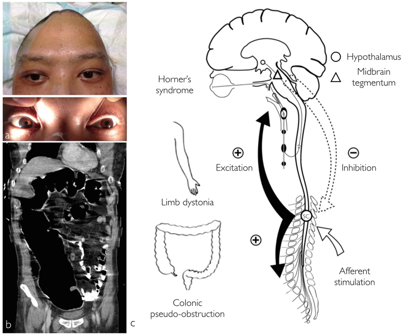
Figure 1. Photographs displaying right side Horner’s syndrome with right hemifacial anhidrosis and left hemihyperhidrosis. Partial eyelid ptosis and meiosis was also observed (a). Acute colonic pseudo-obstruction (b) coronal abdominal computed tomography. Schematic illustrating the pathophysiology behind the concomitant occurrence of central Horner’s syndrome, limb dystonia, colonic pseudo-obstruction and PSH (c). A mesencephalic lesion causes disruption of the hypothalamospinal sympathetic pathway produces Horner’s syndrome and according to the EIR model, loss of normal tonic inhibition of spinal centers from the brainstem (dotted-arrow). Due to this disinhibition minor afferent stimulation (white arrow) elicits spinal allodynia and diffuse spinal cord excitation (black arrow) with PSH (SC, spinal cord center).
Figure 2.
Figure 2. MRI showing evidence of right cerebral peduncle and lateral tegmental injury of the mesencephalon ((a) white arrow, axial T2 sequence; (b) fluid attenuated inversion recovery FLAIR sequence; (c) diffusion-weighted imaging). Injury as a result of downward transtentorial herniation leading to injury of the hypothalamospinal sympathetic tract ((d) white arrowhead, coronal T2; (e) gray arrow, coronal FLAIR; (f) gray arrowhead, sagittal, FLAIR).