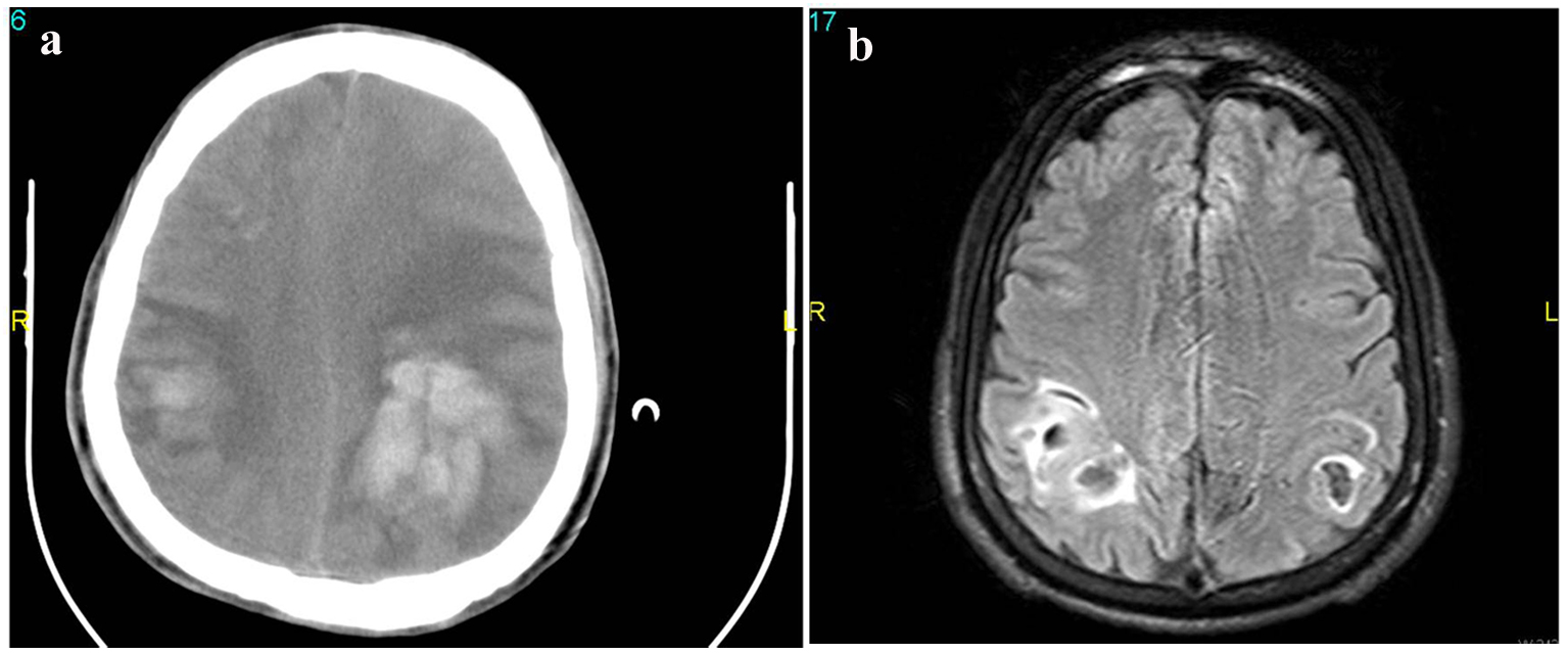
Figure 1. (a) Brain computed tomography of the patient, in the left temporo-parieto-occipital hemorrhagic infarct. (b) Magnetic resonance imaging (MRI) of the patient, in the left temporo-parieto-occipital hemorrhagic infarct.
| Journal of Neurology Research, ISSN 1923-2845 print, 1923-2853 online, Open Access |
| Article copyright, the authors; Journal compilation copyright, J Neurol Res and Elmer Press Inc |
| Journal website http://www.neurores.org |
Case Report
Volume 4, Number 1, February 2014, pages 37-40
A Case Report of Etiology of Cerebral Venous Sinus Thrombosis Developed After Spinal Anesthesia in Asteroid, Doping Using Young Athlete
Figures