Journal of Neurology Research, ISSN 1923-2845 print, 1923-2853 online, Open Access
Article copyright, the authors; Journal compilation copyright, J Neurol Res and Elmer Press Inc
Journal website http://www.neurores.org

Case Report

Volume 3, Number 2, April 2013, pages 73-77


De Novo Dural Arteriovenous Fistula Formation Following Asymptomatic Sinus Thrombosis: A Case Report and Review of the Literature

Figures

Figure 1.
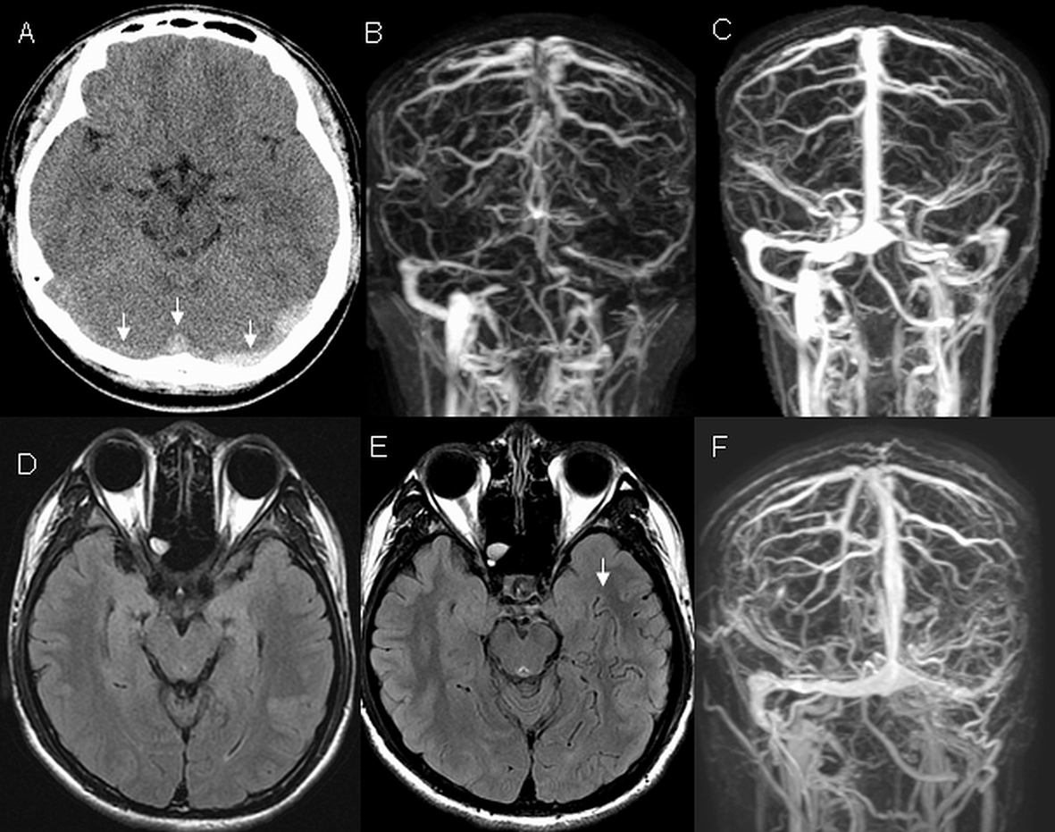
Figure 1. Head imaging. At initial presentation, non-contrast computerized tomography (CT) of the head (A) demonstrated hyperdensities in multiple sinuses including bilateral transverse and superior sagittal sinuses (arrowheads) which presence was verified on magnetic resonance venogram (MRV) (B). After a six-week anticoagulation treatment, repeated MRV (C) showed prominent flow in the superior sagittal and right transverse sinuses. The left transverse sinus recanalized but remained narrow. Flow was also seen in the left sigmoid sinus and left jugular vein. Fluid attenuated inversion recovery (FLAIR) image at six weeks (D) was normal but FLAIR image at 14 months (E) revealed abnormally dilated cerebral vessels representing venous congestion (arrowhead). Follow up MRV at 14 months (F) demonstrated an occlusion of left sigmoid sinus and left jugular vein.
Figure 2.
Figure 2. Digital subtractive angiography. At 14 months, a diagnostic arteriogram demonstrated a DAVF with feeders from the left internal carotid artery (A, arrow indicating the tentorial branch), left external carotid artery (B, arrow indicating occipital branch; arrowhead indicating the patent right transverse sinus), and left vertebral artery (C, arrow indicating the meningeal branch). Prominent cortical venous reflux was also observed (D, arrow).