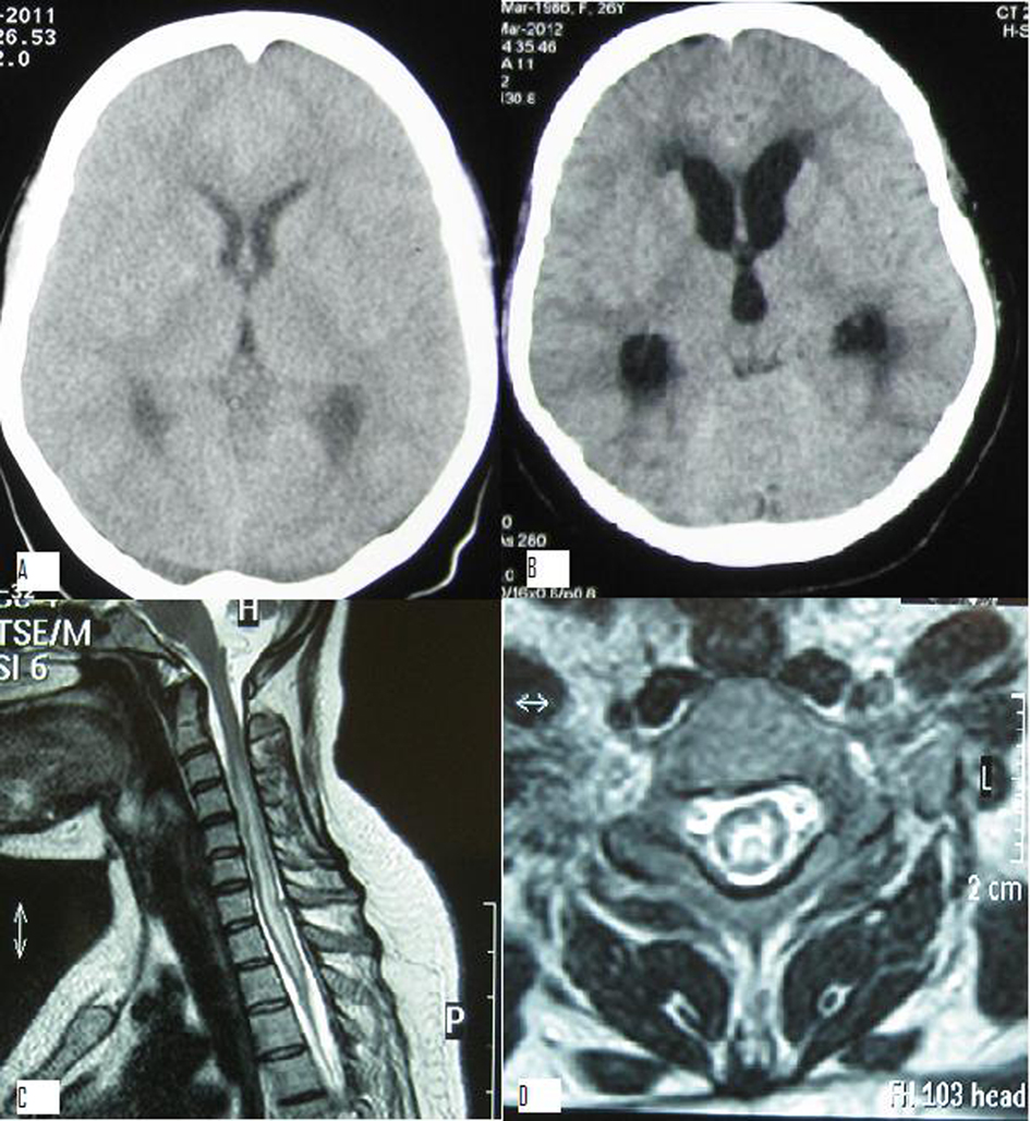
Figure 1. A). Contrast enhanced CT scan head before admission which was normal; B). Contrast enhanced CT scan head done after admission showing communicating hydrocephalus; C). Magnetic resonance imaging (MRI) T2 weighted sagittal image showing intramedullary hyper intensity extending from C3 to T3 level suggestive of syrinx; D). MRI T2 weighted coronal section through C6 level showing intramedullary hyperintensity suggestive of syringomyelia.