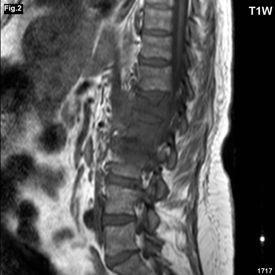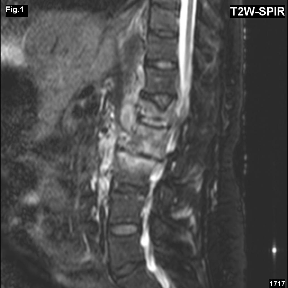
Figure 1. MRI, T2 weighted image with fat suppression (sagittal plane) demonstrated a compression of the vertebral body L1, an increased signal of vertebral bodies L1-3 and in pedicles L1. Intervertebral discs L1/2 and L2/3 were reduced, hyposignal.
| Journal of Neurology Research, ISSN 1923-2845 print, 1923-2853 online, Open Access |
| Article copyright, the authors; Journal compilation copyright, J Neurol Res and Elmer Press Inc |
| Journal website http://www.neurores.org |
Case Report
Volume 3, Number 2, April 2013, pages 78-80
Mycotic Spondylitis Caused by Cladosporium Cladosporides: A Case Report
Figures