


| Journal of Neurology Research, ISSN 1923-2845 print, 1923-2853 online, Open Access |
| Article copyright, the authors; Journal compilation copyright, J Neurol Res and Elmer Press Inc |
| Journal website http://www.neurores.org |
Case Report
Volume 1, Number 3, August 2011, pages 115-116
Creutzfeldt-Jakob Disease: An Atypical Case With Acute Onset and Quick Progression
Adriana Moroa, b, Alisson Pittol Bresciania, Marcus Victor de Oliveiraa, Pedro Henrique de Campos Albinoa, Melina More Bertottia, Rafael Martins Ferreiraa, Luiz Paulo Queiroza, Paulo Mattosinho Filhoa
aNeurology Department, Hospital Governador Celso Ramos, Florianopolis, Santa Catarina, Brazil
bCorresponding author: Adriana Moro, Prefeito Antenor Mesquita 98/101, Centro - Florianopolis - SC, CEP: 88015 - 150
Manuscript accepted for publication August 8, 2011
Short title: Creutzfeldt-Jakob Disease
doi: https://doi.org/10.4021/jnr33w
| Abstract | ▴Top |
Creutzfeldt-Jakob disease (CJD) is a prionic neurodegenerative disease, which leads to patient’s death about 12 months after the onset of symptoms. We report a case of CJD with atypical evolution and unusual findings in MRI.
Keywords: Creutzfeldt-Jakob disease; Dementia; Prionic disease
| Introduction | ▴Top |
Creutzfeldt-Jakob disease (CJD) is a prionic neurodegenerative disease, which leads to patient’s death about 12 months after the onset of symptoms [1, 2]. Patients develop progressive dementia with beginning in the 4th - 5th decade of life, associated with myoclonic jerks, cerebellar syndrome, visual deficits, pyramidal dysfunction or extra-pyramidal acinetic mutism [1]. Additional tests that may aid in diagnosis are brain magnetic resonance imaging (MRI) [3], electroencephalogram (EEG) [4] and protein 14 - 3 - 3 in the cerebrospinal fluid (CSF) [3].
We report a case of CJD with atypical evolution and unusual findings in MRI.
| Case Report | ▴Top |
A 45-year-old previously healthy woman started with abrupt confusion and agitation for the last 24 hours. Eight days later she underwent cranial MRI examination. Restricted diffusion involving the left frontoparietal cortex was observed with further involvement of bilateral fusiform gyri, and a tiny dot of restricted diffusion in the head of the left caudate nucleus (Fig. 1).
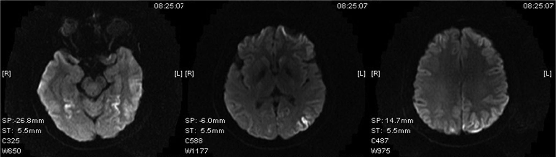 Click for large image | Figure 1. MR diffusion imaging of the brain performed at day 8 shows restricted diffusion in the left frontoparietal cortex in addition to symmetrical involvement of fusiform gyri. A tiny focus of restricted diffusion was observed in the head of the left caudate nucleus. |
At admission, the patient could speak only a few monosyllabic words and obey simple commands. Physical examination showed right hemiparesis with hyperreflexia.
EEG revealed the basic brain electrical activity markedly disorganized and relatively symmetrical, consisting of complex periodic triphasic sharp waves of high amplitude, with regular and diffuse projection and frontocentral predominance, interspersed with background activity slowing and low voltage. In spontaneous drowsiness and sleep, it was observed reduction of the incidence and disappearance of periodic complexes for up to 20 seconds, the background activity replaced by irregular slow delta waves of diffuse projection. With the patient awake and in the presence of repetitive periodic complexes on EEG, the administration of diazepam 10 mg was performed and was observed the disappearance of periodic complexes, even with the patient awake and partially obeying verbal commands, while maintaining expression aphasia. We observed slow, rhythmic, intermittent, high voltage delta activity, of diffuse projection but predominant anterior, characterizing FIRDA.
Routine laboratory tests were normal. CSF analysis was normal, and the test for 14 - 3 - 3 protein was not assessed because it lacked in our service.
At day 21 patient developed akinetic mutism. Progressive worsening of right hemiparesis and myoclonus were observed. The patient died at day 68 owing to acute respiratory failure.
| Discussion | ▴Top |
The CJD should be considered in the differential diagnosis of young patients with rapidly progressive dementia [1, 2].
The World Health Organization criteria for probable sporadic CJD are: progressive dementia; at least two out of the features: myoclonus, visual or cerebellar disturbance, pyramidal/extrapyramidal dysfunction, akinetic mutism; a typical electroencephalogram during an illness of any duration, and/or 14 - 3 - 3 CSF assay with a clinical duration to death less than two years; routine investigations should not suggest an alternative diagnosis [5].
This patient met these criteria. Diffusion WI showed unilateral frontoparietal cortex abnormalities in addition to an involvement of bilateral fusiform gyri. There are no reports of a MRI showing abnormalities so early at the onset of the disease.
The symptoms usually begin in a subacute form and progression to death is inevitable in about 12 months. There are no records of other cases of sporadic CJD with a so abrupt onset and progression to death quickly.
| References | ▴Top |
- Geschwind MD, Haman A, Miller BL. Rapidly progressive dementia. Neurol Clin. 2007;25(3):783-807, vii.
pubmed - Sharma S, Mukherjee M, Kedage V, Muttigi MS, Rao A, Rao S. Sporadic Creutzfeldt-Jakob disease—a review. Int J Neurosci. 2009;119(11):1981-1994.
pubmed doi - Nitrini R, Areza-Fegyveres R, Martins VR, Castro RM, Landemberger MC, Huang N, Bacheschi LA, et al. Asymmetric cortical high signal on diffusion weighted-MRI in a case of Creutzfeldt-Jakob disease. Arq Neuropsiquiatr. 2005;63(2B):519-522.
pubmed doi - Wieser HG, Schindler K, Zumsteg D. EEG in Creutzfeldt-Jakob disease. Clin Neurophysiol. 2006;117(5):935-951.
pubmed doi - Global surveillance, diagnosis and therapy of human transmissible spongiform encephalopathies: Report of a WHO consultation. World Health Organization, Geneva, Switzerland, 9-111 February 1998. www.who.int/emc-documents/tse/docs/whoemczdi989.pdf (Accessed on May 17, 2010).
This is an open-access article distributed under the terms of the Creative Commons Attribution License, which permits unrestricted use, distribution, and reproduction in any medium, provided the original work is properly cited.
Journal of Neurology Research is published by Elmer Press Inc.